 Back
BackThe Cellular Basis of Reproduction and Inheritance: Cell Division, Chromosomes, and Genetic Variation
Study Guide - Smart Notes
 Tailored notes based on your materials, expanded with key definitions, examples, and context.
Introduction to Cell Division and Genetics
Overview of Cell Division and Its Importance
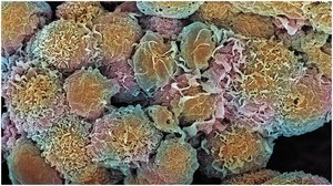
Cell division is a fundamental process in all living organisms, essential for growth, development, maintenance, and reproduction. In multicellular organisms, it enables the formation of tissues and organs, while in unicellular organisms, it is the means of reproduction. Genetic information is faithfully transmitted during cell division, ensuring continuity of life and inheritance of traits.
Cancer arises when mutations disrupt normal cell cycle regulation, leading to uncontrolled cell division and tumor formation.
Clone: An organism or cell produced asexually from one ancestor, to which they are genetically identical.
Chromosome: A structure composed of DNA and proteins that carries genetic information. Humans typically have 46 chromosomes (23 pairs).
Cell Division and Reproduction
Asexual vs. Sexual Reproduction
Organisms reproduce either asexually or sexually, with significant implications for genetic diversity.
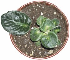
Asexual reproduction: Offspring are produced by a single parent without the involvement of gametes. The offspring are genetically identical to the parent (clones).
Sexual reproduction: Involves the fusion of gametes (egg and sperm), resulting in offspring with unique genetic combinations.
Cell Division in Multicellular Organisms
In multicellular organisms, cell division is responsible for growth, tissue repair, and maintenance. Every cell contains DNA that is a copy of the original zygote formed by the fusion of sperm and egg.
Prokaryotic Cell Division: Binary Fission
Mechanism of Binary Fission
Prokaryotes reproduce asexually through binary fission, a process in which a single cell divides into two genetically identical daughter cells.
Most prokaryotic genes are carried on a single circular DNA molecule (chromosome).
During binary fission, the chromosome is duplicated, and the cell splits into two.
Eukaryotic Chromosomes and the Cell Cycle
Chromosome Structure and Duplication
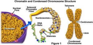
Eukaryotic cells contain multiple, linear chromosomes located in the nucleus. Each chromosome consists of a long DNA molecule with associated proteins, forming chromatin. Chromosomes duplicate before cell division, resulting in two identical sister chromatids joined at the centromere.
Chromatin: Loosely packed DNA and proteins, visible only during cell division as condensed chromosomes.
Sister chromatids: Identical copies of a chromosome, connected at the centromere.
Human Genome
The human genome consists of 46 chromosomes (23 pairs) and approximately 21,000 genes.
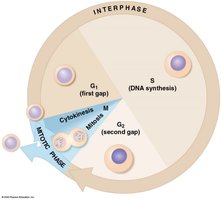
The Cell Cycle
The cell cycle is an ordered sequence of events from the formation of a cell to its own division. It consists of interphase (G1, S, G2) and the mitotic phase (mitosis and cytokinesis).
G1 phase: Cell growth and normal metabolic roles.
S phase: DNA replication (chromosomes are duplicated).
G2 phase: Preparation for cell division.
M phase: Mitosis (nuclear division) and cytokinesis (cytoplasmic division).
Mitosis and Cytokinesis
Phases of Mitosis
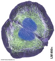
Mitosis is the process by which a eukaryotic cell divides its nucleus, producing two genetically identical daughter nuclei. The phases include:
Prophase: Chromosomes condense, spindle fibers form.
Metaphase: Chromosomes align at the metaphase plate.
Anaphase: Sister chromatids separate to opposite poles.
Telophase: Nuclear envelopes reform around the chromosomes.
Cytokinesis
Cytokinesis divides the cytoplasm, resulting in two separate cells. In animals, a cleavage furrow forms; in plants, a cell plate develops.
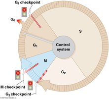
Regulation of the Cell Cycle and Cancer
Cell Cycle Control System
The cell cycle is regulated by checkpoints (G1, G2, M) that ensure proper division. Growth factors are proteins that stimulate cell division by signaling through specific receptors.
If cells do not receive the proper signals at the G1 checkpoint, they may enter a non-dividing state (G0).
Cancer and Tumors
Cancer results from the failure of cell cycle controls, leading to excessive cell division and tumor formation. Tumors can be benign (localized) or malignant (invasive and metastatic). Treatments like radiation and chemotherapy target rapidly dividing cells.
Chromosome Number and Structure
Homologous Chromosomes
Somatic cells contain pairs of homologous chromosomes—one from each parent. Homologous chromosomes carry genes for the same traits at corresponding loci.
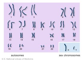
Sex Chromosomes
Humans have two sex chromosomes (XX for females, XY for males) and 22 pairs of autosomes.
Meiosis and Sexual Life Cycles
Meiosis: Reduction Division
Meiosis is the process by which diploid cells produce haploid gametes (eggs and sperm). It involves two consecutive divisions (meiosis I and II) and results in four genetically unique haploid cells.
Meiosis I: Homologous chromosomes separate.
Meiosis II: Sister chromatids separate (similar to mitosis).
Genetic Variation
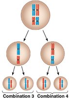
Genetic variation arises from independent assortment of chromosomes, crossing over during prophase I, and random fertilization. In humans, independent assortment and random fertilization can produce about 64 trillion possible combinations in offspring.
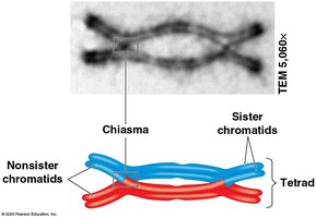
Crossing Over
Crossing over is the exchange of genetic material between nonsister chromatids of homologous chromosomes, resulting in recombinant chromosomes and increased genetic diversity.
Karyotypes and Chromosomal Abnormalities
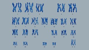
Karyotype Analysis
A karyotype is a photographic inventory of an individual's chromosomes, arranged in homologous pairs. It is used to detect chromosomal abnormalities.
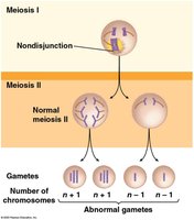
Nondisjunction and Aneuploidy
Nondisjunction is the failure of chromosome pairs or sister chromatids to separate properly during meiosis, leading to gametes with abnormal chromosome numbers. This can result in conditions such as Down syndrome (trisomy 21).
Alterations of Chromosome Structure
Chromosome breakage can lead to structural changes such as deletions, duplications, inversions, and translocations, which may cause genetic disorders or cancer.
Summary Table: Comparison of Mitosis and Meiosis
Feature | Mitosis | Meiosis |
|---|---|---|
Number of divisions | 1 | 2 |
Number of daughter cells | 2 | 4 |
Genetic identity | Identical to parent | Genetically unique |
Chromosome number | Diploid (2n) | Haploid (n) |
Role | Growth, repair, asexual reproduction | Sexual reproduction (gametes) |
Key Terms and Concepts
Somatic cells: Body cells (diploid)
Gametes: Sex cells (haploid)
Diploid (2n): Two sets of chromosomes
Haploid (n): One set of chromosomes
Homologous chromosomes: Chromosome pairs with genes for the same traits
Sister chromatids: Identical copies of a duplicated chromosome
Nondisjunction: Failure of chromosomes to separate properly
Karyotype: Chromosome display for analysis
Equations and Genetic Calculations
Number of possible chromosome combinations in gametes:
(where n = haploid number of chromosomes)
Number of possible combinations in zygote (random fertilization):
For humans (n = 23): combinations per gamete; possible zygote combinations.
