Back
The Genetic Code and Transcription: Study Notes for Genetics Students
Study Guide - Smart Notes
 Tailored notes based on your materials, expanded with key definitions, examples, and context.
The Genetic Code and Transcription
Gene Structure and Function
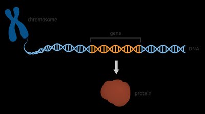
The concept of a gene is central to genetics, representing the basic unit of heredity. Genes are segments of DNA that encode instructions for the synthesis of proteins or functional RNA molecules, such as tRNA and rRNA. These products are responsible for the manifestation of traits in organisms.
Gene: A sequence of DNA that codes for a protein or functional RNA.
Trait: Observable characteristic determined by gene products.
Functional RNA: Includes tRNA (transfer RNA) and rRNA (ribosomal RNA), which are not translated into proteins but play essential roles in protein synthesis.
Protein: The end product of gene expression, responsible for cellular structure and function.
Example: The gene for hemoglobin codes for the protein that carries oxygen in blood.
Transcription and Translation Overview
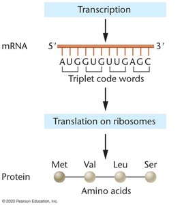
Transcription is the process by which a DNA template strand is used to synthesize messenger RNA (mRNA). Translation follows, where ribosomes decode the mRNA sequence into a chain of amino acids, forming a protein. The genetic code is read in triplets called codons.
Transcription: DNA is copied into mRNA by RNA polymerase.
Translation: Ribosomes read mRNA codons and assemble amino acids into proteins.
Codon: A sequence of three nucleotides in mRNA that specifies an amino acid.
Example: The mRNA sequence AUG codes for methionine, the start amino acid in translation.
Characteristics of the Genetic Code
Structure and Properties
The genetic code is a set of rules by which information encoded in mRNA is translated into proteins. It is composed of codons, each consisting of three ribonucleotides. The code is linear, unambiguous, degenerate, commaless, non-overlapping, and nearly universal.
Linear: mRNA is read from 5' to 3' in a continuous sequence.
Codons: Each codon is a triplet of nucleotides (A, U, G, C).
Unambiguous: Each codon specifies only one amino acid.
Degenerate: Multiple codons can code for the same amino acid.
Commaless: Codons are read sequentially without spaces.
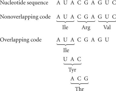
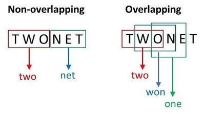
Non-overlapping: Each nucleotide is part of only one codon.
Universal: The code is nearly identical across all living organisms.
Codon Length and Possibilities
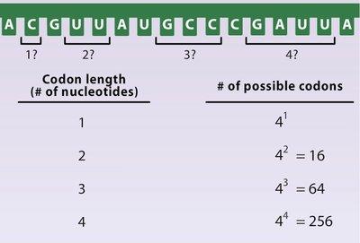
The number of possible codons depends on the length of the codon and the number of nucleotide types. With four nucleotides and a triplet code, there are 64 possible codons.
Formula: where n is the number of nucleotides per codon.
Example: For n = 3, possible codons.
Codon Dictionary
The codon dictionary maps each triplet to its corresponding amino acid. There are specific codons for initiation (start) and termination (stop) of translation.
Start Codon: AUG (methionine)
Stop Codons: UAA, UAG, UGA
Example: UUU codes for phenylalanine, AAA for lysine.
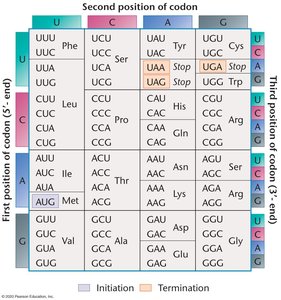
Start and Stop Codons
Translation begins at a start codon and ends at a stop codon. These codons are essential for proper protein synthesis.
Start Codon: Signals the beginning of translation (AUG).
Stop Codons: Signal the end of translation (UAA, UAG, UGA).


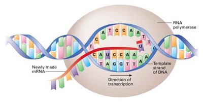
Transcription Mechanism
Transcription is carried out by RNA polymerase, which synthesizes mRNA from the DNA template strand. The direction of transcription is 5' to 3'.
RNA Polymerase: Enzyme responsible for mRNA synthesis.
Template Strand: DNA strand used to make mRNA.
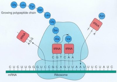
Translation Mechanism
Translation occurs on ribosomes, where tRNA molecules bring amino acids corresponding to each mRNA codon. The polypeptide chain grows as amino acids are added.
Ribosome: Site of protein synthesis.
tRNA: Transfers specific amino acids to the growing polypeptide.
Commaless and Non-Overlapping Code
The genetic code is commaless, meaning codons are read continuously without interruption. It is also non-overlapping, so each nucleotide is part of only one codon.
Commaless: No spaces or punctuation between codons.
Non-overlapping: Each codon is distinct and does not share nucleotides with adjacent codons.
Universality of the Genetic Code
The genetic code is nearly universal, meaning it is conserved across most species. This universality allows for genetic engineering and biotechnology applications.
Universal Code: Most organisms use the same codon assignments.
Example: The same codon specifies the same amino acid in humans, bacteria, and plants.

Experimental Determination of the Genetic Code
Free-Cell System and Radioisotope Labeling
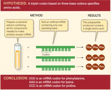
Early experiments used cell-free systems and radioisotope-labeled amino acids to determine which codons specify particular amino acids. Synthetic mRNAs were used to direct protein synthesis in vitro.
Cell-Free System: Mix of ribosomes, tRNAs, amino acids, and synthetic mRNA.
Radioisotope Labeling: Used to track incorporation of specific amino acids.
Trial and Error: Systematic alteration of mRNA sequences to identify codon assignments.
Polynucleotide Phosphorylase and Homopolymers
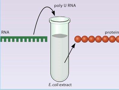
Polynucleotide phosphorylase was used to synthesize mRNA without a DNA template. Homopolymers (runs of the same nucleotide) helped identify codon assignments for specific amino acids.
Homopolymer: mRNA composed of a single nucleotide type (e.g., poly(U)).
Example: Poly(U) directs synthesis of phenylalanine, revealing UUU = Phe.
Heteropolymers and Triplet Binding Assay
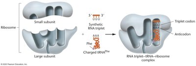
Mixtures of nucleotides (heteropolymers) allowed prediction of codon frequencies and their corresponding amino acids. The triplet binding assay involved synthesizing specific RNA triplets and observing their interaction with tRNA and ribosomes.
Heteropolymer: mRNA with mixed nucleotides.
Triplet Binding Assay: Identifies which tRNA binds to specific RNA triplets in the presence of ribosomes.
Key Findings: The code is degenerate (multiple codons per amino acid) and unambiguous (one codon specifies one amino acid).
Codon Assignments Table
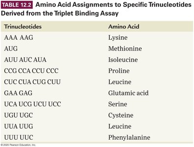
The triplet binding assay produced a table of codon assignments for amino acids. This table is fundamental for understanding translation.
Trinucleotides | Amino Acid |
|---|---|
AAA, AAG | Lysine |
AUG | Methionine |
AUU, AUC, AUA | Isoleucine |
CCG, CCA, CCU, CCC | Proline |
CUC, CUA, CUG, CUU | Leucine |
GAA, GAG | Glutamic acid |
UCA, UCG, UCU, UCC | Serine |
UGU, UGC | Cysteine |
UUG, UUC | Leucine |
UUU, UUC | Phenylalanine |
Wobble Hypothesis
Wobble Position and Codon-Anticodon Pairing
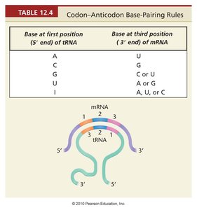
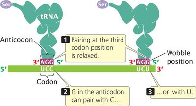
The wobble hypothesis explains how some tRNAs can recognize more than one codon due to relaxed base pairing at the third position of the codon. This contributes to the degeneracy of the genetic code.
Wobble Position: Third base of the codon, allows flexible pairing.
Example: G in the anticodon can pair with C or U in the codon.
Codon-Anticodon Base-Pairing Rules Table
Codon-anticodon pairing rules determine which tRNA can bind to which mRNA codon, especially at the wobble position.
Base at first position (5' end of tRNA) | Base at third position (3' end of mRNA) |
|---|---|
A | U |
C | G |
G | C or U |
U | A or G |
I | A, U, or C |