 Back
BackThe Genetic Code and Translation in Prokaryotes: Structure, Mechanism, and Application
Study Guide - Smart Notes
 Tailored notes based on your materials, expanded with key definitions, examples, and context.
Gene Expression: From DNA to Protein in Prokaryotes
Overview of the Central Dogma
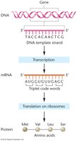
The central dogma of molecular biology describes the flow of genetic information from DNA to RNA to protein. In prokaryotes, transcription and translation are tightly coupled, allowing protein synthesis to begin even before mRNA synthesis is complete.
Transcription: DNA is transcribed into messenger RNA (mRNA).
Translation: Ribosomes read the mRNA sequence to synthesize a polypeptide chain (protein).
Coupling: In prokaryotes, translation can initiate on an mRNA while it is still being transcribed.
Gene Structure in Prokaryotes
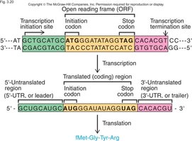
Key Regions of a Bacterial Gene
Bacterial genes are organized into distinct regions that regulate and encode protein synthesis.
Promoter: Upstream regulatory sequence where RNA polymerase binds to initiate transcription (includes –35 and –10 regions).
+1 Site: The transcription start site.
5′ Untranslated Region (5′ UTR): Non-coding region before the start codon; contains the Shine–Dalgarno sequence for ribosome binding.
Open Reading Frame (ORF): Protein-coding region, beginning with the start codon (AUG) and ending with a stop codon (UAA, UAG, UGA).
3′ Untranslated Region (3′ UTR): Non-coding region after the stop codon; may affect mRNA stability and translation efficiency.
Terminator: Sequence signaling the end of transcription.
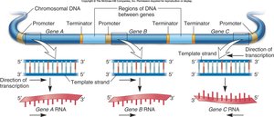
Gene Organization and Transcription
Each gene has its own promoter, coding region, and terminator. Coding regions are separated by noncoding DNA, and transcription produces mRNA complementary to the template strand.
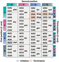
The Genetic Code
Properties of the Genetic Code
The genetic code is a set of rules by which the nucleotide sequence of mRNA is translated into the amino acid sequence of proteins.
Triplet Code: Each codon consists of three nucleotides.
Non-overlapping: Codons are read one after another without overlap.
Degenerate: Multiple codons can specify the same amino acid (redundancy).
Universal: Nearly all organisms use the same code.
Start Codon: AUG (methionine; in bacteria, N-formylmethionine/fMet).
Stop Codons: UAA, UAG, UGA (signal termination of translation).
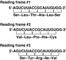
Reading Frames
mRNA can be read in three possible reading frames, but only one is correct for a given gene. The start codon (AUG) establishes the reading frame. Shifting the frame (frameshift mutation) alters the downstream amino acid sequence.
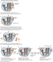
Translation: Mechanism and Stages
Stages of Translation in Prokaryotes
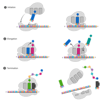
Translation occurs in three main stages: initiation, elongation, and termination.
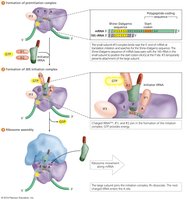
Initiation: Small ribosomal subunit binds the Shine–Dalgarno sequence on mRNA; initiator tRNA (fMet–tRNA) pairs with AUG in the P site; large subunit joins to form the complete ribosome.
Elongation: Charged tRNA enters the A site; peptidyl transferase forms peptide bonds; ribosome translocates one codon; uncharged tRNA exits from the E site.
Termination: Stop codon enters the A site; release factors bind, triggering polypeptide release and ribosome disassembly.
Initiation: The Shine–Dalgarno Sequence
The Shine–Dalgarno (SD) sequence is a purine-rich region in the 5′ UTR of bacterial mRNA. It base-pairs with the 16S rRNA of the small ribosomal subunit, positioning the start codon (AUG) in the P site for accurate initiation.
Typical SD sequence: AGGAGG
Located 5–10 nucleotides upstream of the start codon
Strength of SD–rRNA pairing affects translation efficiency
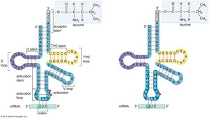
tRNA: The Adaptor Molecule
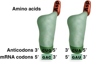
Transfer RNA (tRNA) molecules link mRNA codons to their corresponding amino acids during translation. Each tRNA has a specific anticodon that base-pairs with a codon on the mRNA and carries a specific amino acid at its 3′ end.
Anticodon: Three-base sequence complementary to the mRNA codon.
Aminoacyl-tRNA synthetases: Enzymes that attach the correct amino acid to each tRNA.
Codons and Anticodons
Codons are found on mRNA, while anticodons are found on tRNA. Their base-pairing ensures the correct amino acid is incorporated into the growing polypeptide chain.
Example: If the mRNA codon is 5′–AUG–3′, the tRNA anticodon is 3′–UAC–5′.
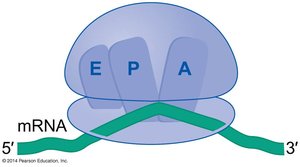
Ribosome Structure and tRNA Binding Sites
The ribosome has three binding sites for tRNA:
A site (Aminoacyl): Entry site for charged tRNA.
P site (Peptidyl): Holds tRNA with the growing polypeptide chain.
E site (Exit): Site where uncharged tRNA exits the ribosome.
Elongation Cycle
During elongation, the ribosome moves along the mRNA in the 5′ to 3′ direction, adding one amino acid per codon. Peptidyl transferase catalyzes peptide bond formation, and translocation shifts the ribosome to the next codon.
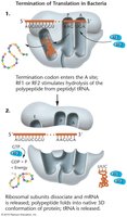
Termination of Translation
Translation ends when a stop codon enters the A site. No tRNA matches these codons; instead, release factors bind, triggering hydrolysis of the bond between the peptide and tRNA, releasing the polypeptide and disassembling the ribosome.
Release Factors: RF1 or RF2 recognize stop codons; RF3 + GTP promote ribosome disassembly.
Practice: Using the Genetic Code
Translating mRNA Sequences
To translate an mRNA sequence, identify the start codon (AUG), read codons in the correct frame, and use the genetic code table to determine the amino acid sequence. Translation stops at the first stop codon encountered.
Example: mRNA 5′–AUG GGC UUU UAA–3′ translates to Met – Gly – Phe – Stop.

Summary Table: Key Features of Prokaryotic Translation
Feature | Description |
|---|---|
Start Codon | AUG (codes for fMet in bacteria) |
Stop Codons | UAA, UAG, UGA |
Shine–Dalgarno Sequence | Purine-rich sequence in 5′ UTR; aligns ribosome with start codon |
tRNA | Adaptor molecule with anticodon and amino acid attachment site |
Ribosome Sites | A (aminoacyl), P (peptidyl), E (exit) |
Release Factors | RF1, RF2, RF3 (terminate translation) |
Additional info:
In prokaryotes, the first methionine is formylated (fMet) to distinguish the start of translation from internal methionines.
Frameshift mutations can result from insertions or deletions that are not multiples of three nucleotides, altering the reading frame and potentially producing nonfunctional proteins.
