 Back
BackThe Molecular Basis of Heredity: Structure and Function of Genetic Material
Study Guide - Smart Notes
 Tailored notes based on your materials, expanded with key definitions, examples, and context.
The Molecular Basis of Heredity, Variation, and Evolution
Criteria for Genetic Material
The genetic material of a cell must fulfill several essential criteria to ensure proper inheritance and function:
Information: Must contain all instructions necessary to build and maintain an organism.
Transmission: Must be reliably passed from parent to offspring.
Replication: Must be capable of accurate copying for inheritance.
Variation: Must allow for changes to account for phenotypic diversity within species.
Historical Determination of Genetic Material
For many years, the identity of the genetic material was unknown. Candidates included lipids, carbohydrates, proteins, and nucleic acids. By the early 1900s, inheritance was linked to chromosomes, which are composed of DNA and protein. Classic experiments established DNA as the genetic material.
Transmission Genetics: Classic Experiments
Griffith's Transformation Experiment
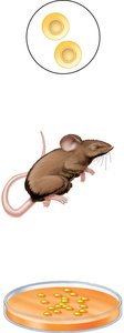
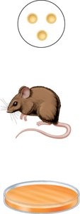
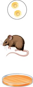
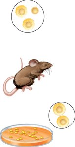
Frederick Griffith studied Streptococcus pneumoniae and discovered transformation, a process where genetic material from dead cells could change the phenotype of living cells.
R strain: No capsule, rough colonies, not protected from immune system.
S strain: Capsule, smooth colonies, protected from immune system.
Key finding: Something from heat-killed S cells transformed R cells into S cells.
Avery, MacLeod, and McCarty Experiment
These scientists fractionated S strain cells into macromolecules and found that only DNA could transform R cells into S cells, confirming DNA as the genetic material.
DNA extract: Transformed R cells.
DNase treatment: Prevented transformation, confirming DNA's role.
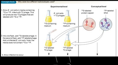
Hershey and Chase Experiment
Studied bacteriophage T2, composed of DNA and protein, to determine which component was the genetic material. Radioactive labeling with 32P (DNA) and 35S (protein) showed that DNA entered the bacterial cell during infection.
Result: Most 32P remained with cells, most 35S was found in the supernatant.
Conclusion: DNA is the genetic material injected into cells.


RNA as Genetic Material in Some Viruses
In 1956, Gierer and Schramm showed that RNA from tobacco mosaic virus (TMV) could cause infection, establishing RNA as the genetic material in some viruses. Many viruses with RNA genomes have since been discovered.
Structure of Nucleic Acids
Levels of Nucleic Acid Structure
DNA and RNA are large macromolecules with several levels of structural complexity:
Nucleotides: The monomeric units.
Strand: Nucleotides linked together.
Double Helix: Two DNA strands interact.
Chromosome: 3D folding and interaction with proteins.

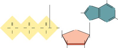
Nucleotide Structure
Nucleotides are composed of a phosphate group, a pentose sugar (deoxyribose in DNA, ribose in RNA), and a nitrogenous base (purine or pyrimidine).
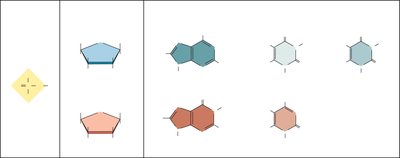
Purines: Adenine (A), Guanine (G) – double ring structure.
Pyrimidines: Cytosine (C), Thymine (T, in DNA), Uracil (U, in RNA) – single ring structure.
Nucleosides and Nucleotides
A nucleoside consists of a base and a sugar. A nucleotide is a nucleoside with one or more phosphate groups.
Adenosine: Adenine + ribose.
Deoxyadenosine: Adenine + deoxyribose.
AMP, ADP, ATP: Adenosine mono-, di-, and triphosphate.
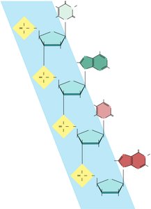
Formation of Nucleic Acid Strands
Nucleotides are covalently linked by phosphodiester bonds, connecting the 5' carbon of one nucleotide to the 3' carbon of another. This gives the strand directionality (5' to 3').
Discovery and Structure of DNA
Scientific Framework for DNA Structure
Many scientists contributed to the discovery of DNA's structure:
Linus Pauling: Proposed alpha-helical structure.
Rosalind Franklin & Maurice Wilkins: Provided x-ray diffraction data.
Erwin Chargaff: Described base pairing rules (A=T, G=C).
Watson & Crick: Compiled data into the double helix model.

Chargaff's Rules
Chargaff found that the amount of adenine equals thymine, and guanine equals cytosine in DNA, supporting the base pairing model.
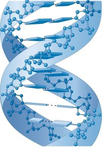
Watson and Crick Model of DNA
DNA consists of two polynucleotide strands held together by hydrogen bonds between bases:
A-T: 2 hydrogen bonds.
G-C: 3 hydrogen bonds.
Bases: Located on the interior, stacked like plates.
Sugar-phosphate backbone: On the exterior.

Key Features of DNA Double Helix
Antiparallel strands: One runs 5' to 3', the other 3' to 5'.
Right-handed helix: Spirals clockwise.
10 bases per turn: Each complete twist is 3.4 nm.
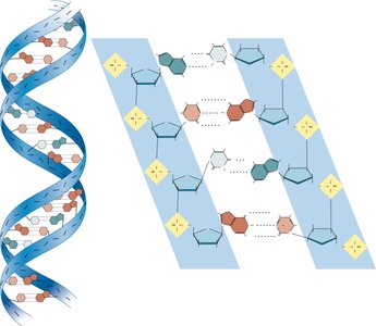
Major and Minor Grooves
DNA has two asymmetrical grooves on its surface: the major and minor grooves. Proteins often bind to the major groove, which can be sequence-specific.
Alternative DNA Structures
DNA can adopt different secondary structures:
B-DNA: Predominant form in cells.
A-DNA: Found in vitro under low humidity, 11 bp per turn.
Z-DNA: Left-handed helix, favored by GG-rich sequences and high salt; may play roles in transcription and recombination.


Triplex DNA
Triplex DNA can form when a synthetic strand binds to the major groove of double-stranded DNA. This structure is sequence-specific and can affect replication, transcription, and recombination.
RNA Structure
Primary and Secondary Structure of RNA
RNA is usually single-stranded but can form short double-stranded regions through complementary base pairing (A-U, C-G). RNA double helices are right-handed and typically have the A form with 11-12 base pairs per turn.
Secondary structures: Hairpins, bulge loops, internal loops, multibranched junctions.
Factors influencing structure: Base pairing, base stacking, interactions with ions, small molecules, and proteins.
Transfer RNA (tRNA) Structure
tRNAs fold into a characteristic shape, carrying amino acids at the 3' end and exposing the anticodon region for interaction with mRNA during translation.
Summary Table: DNA vs. RNA
Feature | DNA | RNA |
|---|---|---|
Sugar | Deoxyribose | Ribose |
Base | A, T, G, C | A, U, G, C |
Strandedness | Double-stranded | Single-stranded (usually) |
Function | Genetic material | Protein synthesis, regulation, genetic material in some viruses |
Additional info: The notes expand on brief points with academic context, definitions, and examples to ensure completeness and clarity for genetics students.
