 Back
The Molecular Biology of Translation: Study Notes for Genetics Students
Study Guide - Smart Notes
 Tailored notes based on your materials, expanded with key definitions, examples, and context.
The Molecular Biology of Translation
Polypeptides and Amino Acids
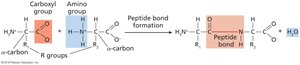
Polypeptides are linear chains of amino acids assembled at ribosomes, forming the primary structure of proteins. The distinctive features of amino acids, such as their side chains (R-groups), determine their chemical properties and interactions.
Amino acids are the building blocks of polypeptides; there are 20 common amino acids.
Covalent peptide bonds link amino acids together.
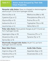
Amino acids can be classified as nonpolar, polar, or electrically charged based on their side chains.
Polypeptide and Transcript Structure
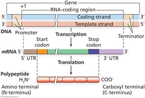
Ribosomes translate messenger RNA (mRNA) in the 5' to 3' direction, assembling amino acids in the order specified by codons. The mRNA sequence determines the resulting amino acid sequence, with translation boundaries defined by start and stop codons.
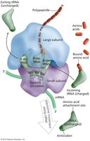
Ribosomes are complexes of rRNAs and proteins that facilitate translation.
The 5' untranslated region (UTR) and 3' UTR are segments of mRNA outside the translated region.
Polypeptide Structure
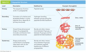
Polypeptides exhibit four levels of structural organization, each stabilized by specific interactions.
Primary structure: Sequence of amino acids.
Secondary structure: Alpha helices and beta-pleated sheets formed by hydrogen bonding.
Tertiary structure: Three-dimensional folding due to interactions among R-groups.
Quaternary structure: Association of multiple polypeptide chains.
Ribosome Structure and Function
Ribosome Composition
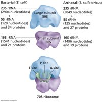
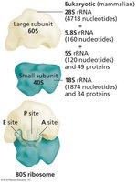
Ribosomes are composed of two subunits, each containing rRNA and proteins. The composition and size differ between bacteria, archaea, and eukaryotes, measured in Svedberg units (S).
Bacterial ribosomes: 30S (small) + 50S (large) = 70S
Eukaryotic ribosomes: 40S (small) + 60S (large) = 80S
Archaeal ribosomes: Similar rRNA to bacteria, but different protein composition.
Functional Sites of Ribosomes
Ribosomes contain three important sites:
P site (peptidyl): Holds tRNA with attached polypeptide.
A site (aminoacyl): Binds incoming tRNA with amino acid.
E site (exit): Allows tRNA to exit after amino acid transfer.
Phases of Translation
Initiation
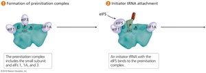
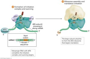
Translation initiation involves assembly of the ribosome at the start codon, aided by initiation factors and GTP hydrolysis. In eukaryotes, the small subunit scans the mRNA for the start codon, typically the first AUG embedded in the Kozak sequence.
Initiator tRNA binds the start codon.
Initiation factors (eIFs) regulate ribosome assembly.
Kozak sequence: 5'-ACCAUGG-3' consensus for start codon recognition.
Elongation
Elongation is the process of sequential addition of amino acids to the growing polypeptide chain, facilitated by elongation factors and GTP hydrolysis.
Charged tRNAs are recruited to the A site.
Peptide bonds are formed between amino acids.
The ribosome translocates along the mRNA.
Termination
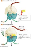
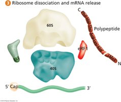
Termination occurs when a stop codon enters the A site. Release factors bind the stop codon, releasing the polypeptide and dissociating the ribosome.
Stop codons: UAA, UAG, UGA.
Single release factor (eRF1) in eukaryotes recognizes all stop codons.
Translation Efficiency and Polyribosomes
Polyribosomes
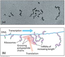
Translation is highly efficient, with multiple ribosomes (polyribosomes) simultaneously translating a single mRNA molecule.
Bacterial cells contain ~20,000 ribosomes.
Polyribosomes increase protein synthesis efficiency.
Coupling of Transcription and Translation
In bacteria, transcription and translation are coupled, allowing ribosomes to translate mRNA as it is being synthesized. In eukaryotes, mRNA is processed in the nucleus before translation in the cytoplasm.
Polycistronic vs. Monocistronic mRNA
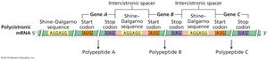
Bacterial and archaeal operons produce polycistronic mRNAs, encoding multiple proteins. Eukaryotic mRNAs are typically monocistronic, encoding a single polypeptide.
The Genetic Code
Codons and the Triplet Code
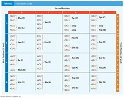
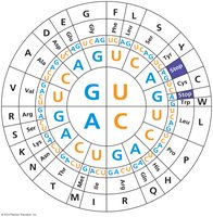
The genetic code is the correspondence between mRNA codons and amino acids. Each codon consists of three nucleotides, and the code is redundant, with multiple codons specifying the same amino acid.
64 codons: 61 specify amino acids, 3 are stop codons.
Synonymous codons specify the same amino acid.
Third-Base Wobble
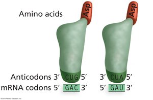
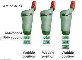
Third-base wobble refers to flexible pairing at the third position of the codon, allowing fewer tRNA molecules to recognize multiple codons.
Isoaccepting tRNAs recognize synonymous codons.
Wobble pairing increases efficiency of translation.
Universality and Exceptions of the Genetic Code
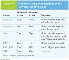
The genetic code is nearly universal across organisms, with a few exceptions found in mitochondria and some protozoa.
tRNA Charging and Protein Processing
Charging tRNA Molecules
tRNAs are charged with their corresponding amino acids by aminoacyl-tRNA synthetases, ensuring fidelity in translation.
Protein Folding and Posttranslational Processing
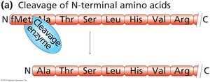
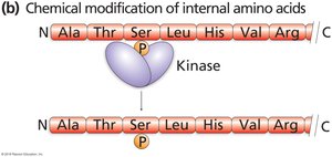
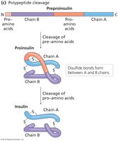
Proteins undergo folding and posttranslational modifications to achieve their functional forms. These modifications include cleavage, phosphorylation, methylation, and addition of carbohydrate side chains.
Removal of N-terminal amino acids is common.
Phosphorylation by kinases can activate or inactivate proteins.
Polypeptide cleavage produces functional proteins, such as insulin.
Signal Hypothesis and Protein Sorting
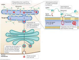
Signal sequences direct proteins to their cellular destinations, such as the endoplasmic reticulum (ER) and Golgi apparatus, for sorting and secretion.
Experimental Deciphering of the Genetic Code
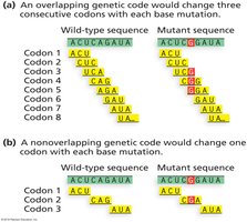
No Overlap and Triplet Nature of the Code
Experiments demonstrated that codons do not overlap and are read continuously, with no gaps. Frameshift mutations alter the reading frame, affecting all downstream codons.
Deciphering Codon Assignments
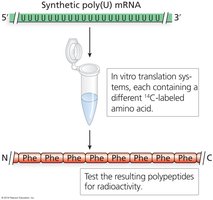
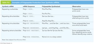
Experiments by Nirenberg, Matthaei, and Khorana used synthetic mRNAs to determine which codons specify which amino acids.
Poly(U) mRNA produces polypeptides of phenylalanine (Phe).
Repeating di-, tri-, and tetranucleotides helped define codon assignments.
Final Decoding Experiments
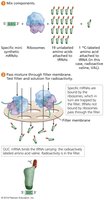
Nirenberg and Leder used mini-RNAs to test all 64 codons, identifying their corresponding amino acids and the three stop codons.
Summary Table: Polypeptide Structure
Level | Description | Stabilized by | Example |
|---|---|---|---|
Primary | Sequence of amino acids | Peptide bonds | Hemoglobin (one α-helix) |
Secondary | α-helices and β-sheets | Hydrogen bonding | Hemoglobin (one α-helix) |
Tertiary | 3D folding | Bonds/interactions among R-groups | Hemoglobin (one subunit) |
Quaternary | Multiple polypeptides | Bonds/interactions among subunits | Hemoglobin (four polypeptide subunits) |
Summary Table: Amino Acids Grouped by Side Chain Properties
Type | Amino Acids |
|---|---|
Nonpolar | Ala, Gly, Ile, Leu, Met, Phe, Pro, Trp, Val |
Polar | Asn, Cys, Gln, Ser, Thr, Tyr |
Electrically charged | Asp, Glu, Arg, His, Lys |
Summary Table: Genomes Using Modifications of the Universal Genetic Code
Codon | Universal Code | Unusual Code | Genome |
|---|---|---|---|
AGA, AGG | Arg | Stop | Mitochondria in plants, animals, yeast |
AUA, AUU | Ile | Met | Mitochondria in plants, animals, yeast |
UGA | Stop | Trp | Mitochondria in plants, animals, yeast, Mycoplasma |
CUN | Leu | Thr | Mitochondria in yeast |
UAA, UAG | Stop | Gln | Green algae, protozoa |
UGA | Stop | Cys | Protozoa |
Key Equations
Peptide bond formation:
Number of possible codons:
