 Back
BackTranscription and RNA Processing: From DNA to Functional RNA
Study Guide - Smart Notes
 Tailored notes based on your materials, expanded with key definitions, examples, and context.
RNA Structure and Types
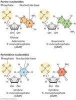
The Four RNA Ribonucleotides
RNA is composed of four ribonucleotides, each consisting of a ribose sugar, a phosphate group, and a nitrogenous base. These bases are divided into purines (adenine and guanine) and pyrimidines (cytosine and uracil). The structure of these nucleotides is fundamental to RNA's role in genetic information transfer and protein synthesis.
Purine nucleotides: Adenosine (AMP) and Guanosine (GMP)
Pyrimidine nucleotides: Uridine (UMP) and Cytidine (CMP)
RNA Assembly and Structure
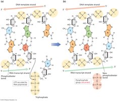
RNA strands are assembled by the formation of phosphodiester bonds between adjacent nucleotides, creating a sugar-phosphate backbone similar to DNA. RNA is synthesized from a DNA template using complementary base pairing, where adenine pairs with uracil and cytosine pairs with guanine.
Categories of RNA
There are several categories of RNA, each with distinct functions:
Messenger RNA (mRNA): Short-lived intermediaries between DNA and protein synthesis.
Ribosomal RNA (rRNA): Combines with proteins to form ribosomes, the site of protein synthesis.
Transfer RNA (tRNA): Binds amino acids and brings them to the ribosome for protein assembly.
Functional RNAs: Includes rRNA, tRNA, telomerase RNA, small nuclear RNA (snRNA), microRNA (miRNA), and small interfering RNA (siRNA), which play roles in gene regulation, RNA processing, and chromosome maintenance.
Bacterial Transcription: Mechanism and Regulation
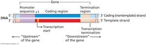
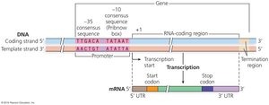
Gene Structure and Transcriptional Units
A gene consists of several functional segments:
Promoter: Located upstream (5') of the transcription start site (+1), controls RNA polymerase access.
Coding region: Contains the information for protein synthesis.
Termination region: Regulates the cessation of transcription, located downstream (3') of the coding region.
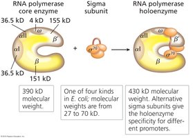
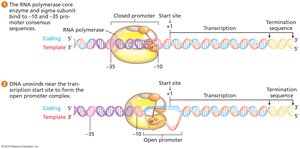
Bacterial RNA Polymerase and Sigma Factor
Bacterial RNA polymerase is a multi-subunit enzyme responsible for synthesizing all types of RNA in bacteria. The core enzyme consists of five subunits (two α, two β, and one ω). The sigma (σ) subunit associates with the core to form the holoenzyme, which is required for promoter recognition and initiation of transcription.
Promoter Recognition and Consensus Sequences
Promoters are double-stranded DNA sequences that serve as binding sites for RNA polymerase and other transcription proteins. In bacteria, two key consensus sequences are found:
-10 region (Pribnow box): 5'-TATAAT-3'
-35 region: 5'-TTGACA-3'
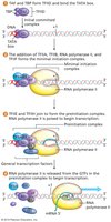
Stages of Bacterial Transcription
Transcription in bacteria occurs in four stages:
Promoter recognition: RNA polymerase holoenzyme binds to the promoter.
Initiation: Formation of the closed promoter complex, followed by DNA unwinding to form the open promoter complex.
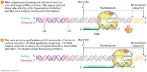
Elongation: RNA synthesis begins at the +1 site; the sigma subunit dissociates after the first 8-10 nucleotides are joined.
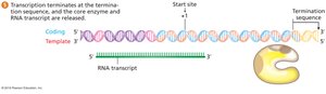
Termination: RNA polymerase encounters a termination sequence and releases the RNA transcript.
Transcription Termination Mechanisms
There are two main mechanisms for terminating transcription in bacteria:
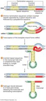
Intrinsic (Rho-independent) termination: Involves formation of a hairpin loop in the RNA transcript, followed by a string of uracils, causing the RNA polymerase to dissociate.
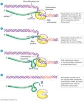
Rho-dependent termination: Requires the rho protein, which binds to the rut site on the RNA and moves toward the polymerase, causing release of the transcript.
Eukaryotic Transcription: Complexity and Regulation
Eukaryotic RNA Polymerases
Eukaryotes possess three distinct RNA polymerases:
RNA polymerase I: Transcribes rRNA genes.
RNA polymerase II: Transcribes protein-coding genes and most snRNA genes.
RNA polymerase III: Transcribes tRNA genes, one snRNA gene, and one rRNA gene.
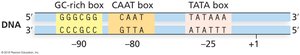
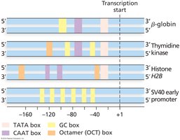
Eukaryotic Promoter Elements
Eukaryotic promoters are more variable than bacterial promoters and contain several consensus sequence elements:
TATA box (Goldberg–Hogness box): Located at about -25, consensus 5'-TATAAA-3'
CAAT box: Often found near -80
GC-rich box: Consensus 5'-GGGCGG-3', may be at -90 or further upstream
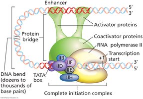
Transcription Initiation in Eukaryotes
Initiation of transcription by RNA polymerase II requires the assembly of general transcription factors (TFII proteins) at the promoter. The TATA-binding protein (TBP) within TFIID binds the TATA box, followed by the sequential addition of other factors to form the preinitiation complex (PIC), which positions RNA polymerase II at the +1 site.
Enhancer and Silencer Sequences
Enhancers are DNA elements that increase transcription levels by binding activator proteins, which interact with the transcription complex at the promoter via DNA bending. Silencers are DNA elements that repress transcription by binding proteins that inhibit the transcription machinery.
Posttranscriptional Processing in Eukaryotes
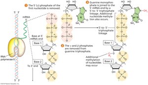
5′ Capping
After the first 20–30 nucleotides of mRNA are synthesized, a guanine is added to the 5′ end by guanylyl transferase, followed by methylation. This 5′ cap protects mRNA from degradation, aids in nuclear export, splicing, and translation initiation.
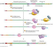
3′ Polyadenylation
The 3′ end of pre-mRNA is cleaved and a poly-A tail is added by polyadenylate polymerase. This modification enhances mRNA stability, export, and translation.


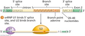
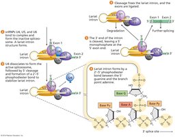
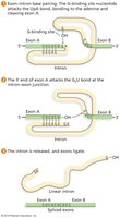
Intron Splicing
Introns are non-coding sequences removed from pre-mRNA by the spliceosome, a complex of snRNAs and proteins. Splicing requires precise recognition of the 5′ splice site (GU), branch site (with an invariant adenine), and 3′ splice site (AG). Errors in splicing can lead to aberrant proteins.

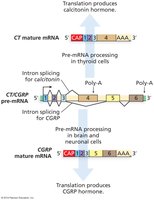
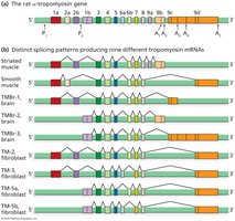
Alternative Splicing and Processing
Alternative splicing allows a single gene to produce multiple mRNA variants and thus different proteins. This process is regulated by cell type and developmental stage, greatly expanding proteomic diversity.
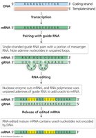
Self-Splicing Introns and RNA Editing
Some introns can self-splice without the spliceosome, acting as ribozymes. RNA editing can alter nucleotide sequences posttranscriptionally, such as by insertion, deletion, or base substitution, further diversifying gene expression.
Translation: From mRNA to Protein
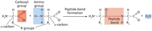
Amino Acid Structure and Peptide Bond Formation
Proteins are polymers of amino acids linked by peptide bonds. Each amino acid contains a central carbon, an amino group, a carboxyl group, and a unique R group. Peptide bonds form between the carboxyl group of one amino acid and the amino group of the next, releasing water.
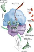
Ribosome Structure and Function
Ribosomes are composed of rRNA and proteins, with distinct large and small subunits. They facilitate the decoding of mRNA and the synthesis of polypeptides. Key sites include the A (aminoacyl), P (peptidyl), and E (exit) sites.
Translation Initiation, Elongation, and Termination
Translation proceeds in three phases:
Initiation: Ribosome assembly at the start codon, aided by initiation factors.
Elongation: Sequential addition of amino acids, facilitated by elongation factors and GTP hydrolysis.
Termination: Release factors recognize stop codons, releasing the completed polypeptide.
The Genetic Code and Third-Base Wobble
The genetic code is a triplet code, with 64 codons specifying 20 amino acids and three stop signals. Third-base wobble allows some tRNAs to recognize multiple codons, increasing translation efficiency.
Charging tRNA Molecules
Aminoacyl-tRNA synthetases attach the correct amino acid to each tRNA, ensuring fidelity in translation. ATP provides the energy for this process.
Protein Folding and Posttranslational Processing
After translation, polypeptides fold into functional structures and may undergo modifications such as cleavage, phosphorylation, or addition of chemical groups. Signal sequences direct proteins to their cellular destinations.
Additional info: This guide covers the molecular biology of transcription and translation, including RNA structure, transcription mechanisms in prokaryotes and eukaryotes, RNA processing, and the translation of mRNA into protein. It is suitable for exam preparation in a college-level genetics course.
