 Back
BackTranslation and Post-Translational Modifications: From mRNA to Functional Protein
Study Guide - Smart Notes
 Tailored notes based on your materials, expanded with key definitions, examples, and context.
Translation: Decoding the Genetic Message
Overview of Translation
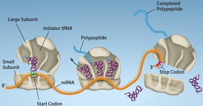
Translation is the process by which the genetic information encoded in messenger RNA (mRNA) is used to synthesize a specific polypeptide (protein). This process occurs in the cytoplasm and is facilitated by ribosomes, transfer RNAs (tRNAs), and various protein factors. Translation is a central event in gene expression, linking the nucleotide sequence of genes to the amino acid sequence of proteins.
Key Components: mRNA, tRNA, ribosomes, amino acids, and translation factors.
Stages: Initiation, elongation, and termination.
Genetic Code: The sequence of nucleotides in mRNA is read in sets of three (codons), each specifying an amino acid.
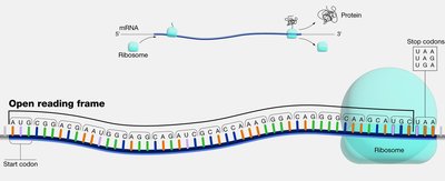
Open Reading Frame (ORF)
An open reading frame (ORF) is a continuous stretch of codons in an mRNA that begins with a start codon (usually AUG) and ends with a stop codon (UAA, UAG, or UGA). The ORF defines the region that will be translated into a protein.
Start Codon: Typically AUG, coding for methionine.
Stop Codons: UAA, UAG, UGA; signal the end of translation.
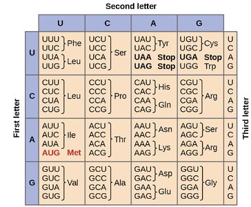
The Genetic Code and Codon Table
The genetic code is degenerate, meaning that most amino acids are specified by more than one codon. The first and second bases of the codon are most important for determining the amino acid, while the third base (the "wobble" position) is often less critical.
64 possible codons (43 combinations) encode 20 amino acids and three stop signals.
Wobble Hypothesis: Flexibility in base pairing at the third codon position allows some tRNAs to recognize multiple codons.
Translation Machinery
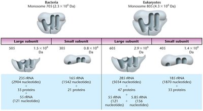
Ribosomes
Ribosomes are large ribonucleoprotein complexes composed of ribosomal RNA (rRNA) and proteins. They consist of two subunits (large and small) and provide the site for mRNA decoding and peptide bond formation.
Prokaryotic Ribosomes: 70S (50S large + 30S small subunits)
Eukaryotic Ribosomes: 80S (60S large + 40S small subunits)
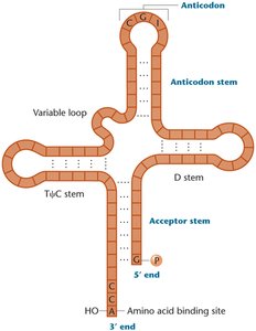
Transfer RNAs (tRNAs)
tRNAs are small, stable RNA molecules (75–90 nucleotides) that serve as adaptors, matching amino acids to their corresponding codons in the mRNA. Each tRNA has a specific anticodon and an acceptor stem for amino acid attachment.
Cloverleaf Structure: Four stems and three loops, with the anticodon loop recognizing the mRNA codon.
3' CCA Tail: Site of amino acid attachment.
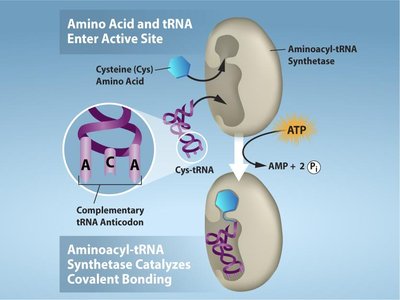
Charging of tRNAs
Aminoacyl-tRNA synthetases are enzymes that attach the correct amino acid to its corresponding tRNA, a process requiring ATP. There are 20 different synthetases, one for each amino acid, ensuring high specificity.
Reaction: Amino acid + tRNA + ATP → aminoacyl-tRNA + AMP + PPi
Importance: Accurate charging is essential for correct translation.
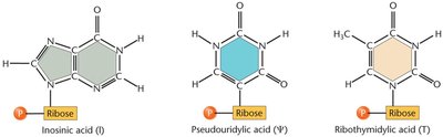
tRNA Modifications
tRNAs undergo extensive post-transcriptional modifications, including base modifications, trimming of precursor sequences, addition of the CCA tail, and removal of introns. These modifications enhance tRNA stability, accuracy, and flexibility in codon recognition.
Modified Bases: Inosine, pseudouridine, ribothymidine, etc.
Unique Structure: Ensures proper function during translation.
Ribosomal DNA (rDNA) and rRNA Genes
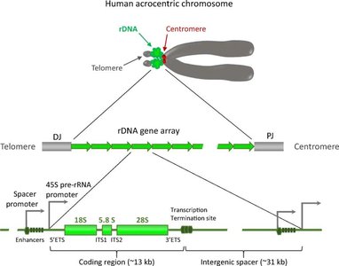
Organization of rDNA
Ribosomal DNA (rDNA) encodes rRNA molecules and is organized in tandem repeats within the genome. In humans, these repeats are found on the short arms of acrocentric chromosomes (13, 14, 15, 21, 22) and are separated by noncoding spacer DNA.
rDNA Clusters: Located in nucleolus organizer regions (NORs).
Repeat Units: Each contains coding regions for 18S, 5.8S, and 28S rRNAs, separated by internal and external transcribed spacers (ITS, ETS).
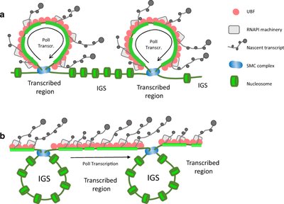
Spatial Organization of rDNA
Transcriptionally active rDNA repeats form loops that facilitate efficient transcription and recycling of RNA polymerase I. Inactive regions may be looped out, allowing for regulation of rRNA synthesis.
SMC Complexes: Organize and stabilize chromosomal regions.
UBF: Upstream binding factor, a transcription factor for rDNA.
Mechanism of Translation
Initiation (Prokaryotes)
In bacteria, translation initiation involves the small ribosomal subunit binding to the Shine–Dalgarno sequence on the mRNA, positioning the start codon (AUG) in the P site. The initiator tRNA (fMet-tRNAfMet) binds, and initiation factors (IF1, IF2, IF3) assist in complex formation. The large subunit then joins, and elongation begins.
Elongation (Bacteria)
During elongation, aminoacyl-tRNAs enter the A site, peptide bonds are formed, and the ribosome translocates along the mRNA. Elongation factors (EF-Tu, EF-G) facilitate these steps, and the process repeats until a stop codon is reached.
Polysomes: Multiple ribosomes can translate a single mRNA simultaneously.
Termination
Termination occurs when a stop codon enters the A site. Release factors (RF1, RF2, RF3) recognize the stop codon, promote hydrolysis of the polypeptide from the tRNA, and disassemble the translation complex.
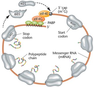
Translation in Eukaryotes
Eukaryotic translation is more complex, involving larger ribosomes, more initiation factors, and additional mRNA processing (5' cap, poly-A tail). The Kozak sequence surrounds the start codon, enhancing initiation efficiency. Ribosomes assemble at the cap, scan for the start codon, and translation proceeds in the cytoplasm.
One-Gene–One-Enzyme and One-Gene–One-Polypeptide Hypotheses
Historical Perspective
The one-gene–one-enzyme hypothesis, proposed by Beadle and Tatum, established that each gene encodes a specific enzyme. This concept evolved into the one-gene–one-polypeptide hypothesis, reflecting the fact that not all proteins are enzymes and some proteins are composed of multiple polypeptide chains.
Experimental Evidence: Mutant strains of Neurospora requiring specific supplements revealed gene-enzyme relationships.
Protein Folding, Modification, and Targeting
Protein Folding
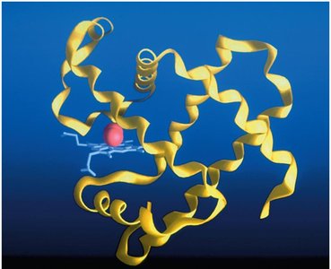
After translation, polypeptides fold into specific three-dimensional structures, often with the assistance of chaperone proteins. Proper folding is essential for biological function; misfolding can lead to disease.
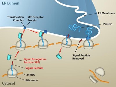
Signal Sequences: Short amino-terminal sequences direct proteins to specific cellular compartments (e.g., ER, mitochondria).
Chaperones: Assist in correct folding and prevent aggregation.
Protein Structure Levels
Proteins exhibit four levels of structure:
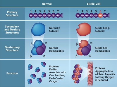
Primary: Linear sequence of amino acids.
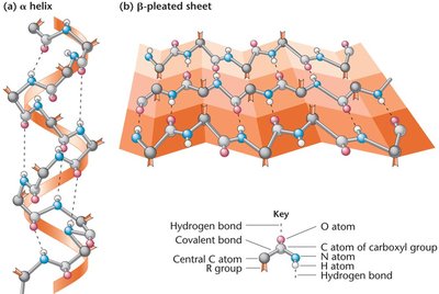
Secondary: Local folding into α-helices and β-pleated sheets.
Tertiary: Overall three-dimensional conformation.
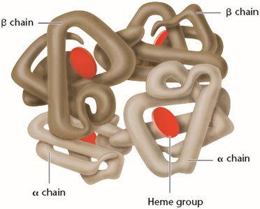
Quaternary: Association of multiple polypeptide chains.
Amino Acids and Peptide Bonds
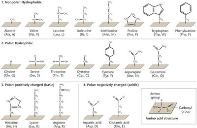
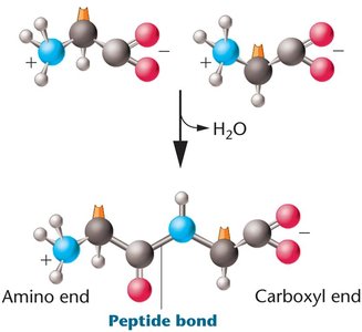
Amino acids are the building blocks of proteins, each with a central carbon, amino group, carboxyl group, and variable R group. Peptide bonds form via dehydration reactions between the carboxyl group of one amino acid and the amino group of another.
Mutations and Protein Function
Effects of Mutations
Mutations in the coding sequence can alter the amino acid sequence of proteins, potentially affecting their structure and function. For example, insertion mutations can shift the reading frame (frameshift), leading to dramatic changes in the protein product.
Sickle Cell Anemia: A point mutation in the β-globin gene changes glutamic acid to valine, causing hemoglobin molecules to aggregate and red blood cells to adopt a sickle shape.
Post-Translational Modifications (PTMs)
Types and Functions of PTMs
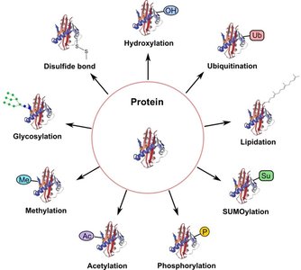
After translation, proteins often undergo covalent modifications that regulate their activity, stability, localization, and interactions. These modifications are crucial for the functional diversity of the proteome.
Modification | Main Function |
|---|---|
Phosphorylation | Regulates enzyme activity and cell signaling |
Acetylation | Controls gene expression and protein stability |
Methylation | Modulates protein interactions and gene regulation |
Glycosylation | Aids protein folding, stability, and cell recognition |
Ubiquitination | Tags proteins for degradation by the proteasome |
SUMOylation | Regulates protein localization and transcription |
Lipidation | Targets proteins to cell membranes |
Hydroxylation | Important for collagen stability and oxygen sensing |
Disulfide bonds | Stabilize protein structure |
Protein Domains and Functional Diversity
Protein Domains
Protein domains are distinct structural and functional units within a protein, typically 50–300 amino acids in length. Different domains confer specific functional capabilities, such as DNA binding, dimerization, or ligand binding.
Transcription Factors: Modular proteins with DNA-binding and effector domains.
Protein-Protein Interactions: Induce conformational changes essential for function and signaling.
Regulation of Gene Expression
Multiple Levels of Regulation
Gene expression is regulated at various stages, including chromatin accessibility, transcription, mRNA processing, translation, and protein degradation. The final protein level depends on both synthesis and degradation rates.
Ubiquitination: Marks proteins for degradation by the proteasome, controlling protein longevity and gene expression.
Other PTMs: Phosphorylation, cleavage, and translocation also regulate protein activity and gene expression.
