 Back
BackTranslation and Post-Translational Modifications in Genetics
Study Guide - Smart Notes
 Tailored notes based on your materials, expanded with key definitions, examples, and context.
Translation: Decoding the Genetic Message
Overview of Translation
Translation is the process by which the genetic information encoded in messenger RNA (mRNA) is used to synthesize polypeptides (proteins). This process occurs in the cytoplasm and is fundamental to gene expression, linking the nucleotide sequence of genes to the amino acid sequence of proteins.
Key Components: mRNA, transfer RNA (tRNA), and ribosomes.
Stages: Initiation, elongation, and termination.
Genetic Code: The sequence of nucleotide triplets (codons) in mRNA determines the sequence of amino acids in the protein.
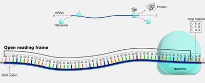
Open Reading Frame (ORF)
An open reading frame (ORF) is a continuous stretch of codons in an mRNA that begins with a start codon (usually AUG) and ends with a stop codon (UAA, UAG, or UGA). The ORF defines the region that can be translated into a protein.
Start Codon: Typically AUG, coding for methionine.
Stop Codons: UAA, UAG, UGA; signal the end of translation.
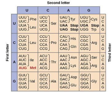
The Genetic Code
The genetic code is a set of rules by which the sequence of nucleotides in mRNA is translated into the sequence of amino acids in a protein. It is nearly universal and degenerate, meaning that most amino acids are encoded by more than one codon.
Wobble Position: The third base of the codon is less important for specifying the amino acid, allowing for some flexibility (wobble hypothesis).
Degeneracy: 64 possible codons but only 20 amino acids.
Translation Machinery
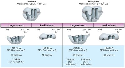
Ribosomes
Ribosomes are large ribonucleoprotein complexes that catalyze protein synthesis. They consist of two subunits (large and small) and are composed of ribosomal RNA (rRNA) and proteins.
Prokaryotic Ribosomes: 70S (50S large + 30S small subunit)
Eukaryotic Ribosomes: 80S (60S large + 40S small subunit)
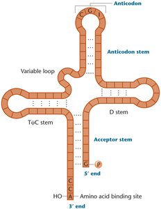
Transfer RNAs (tRNAs)
tRNAs are small, stable RNA molecules that serve as adapters, matching amino acids to their corresponding codons in the mRNA during translation. Each tRNA has an anticodon that pairs with the mRNA codon and an acceptor stem for amino acid attachment.
Structure: Cloverleaf secondary structure with four stems and three loops.
Anticodon Loop: Contains the anticodon sequence complementary to the mRNA codon.
3' CCA End: Site of amino acid attachment.
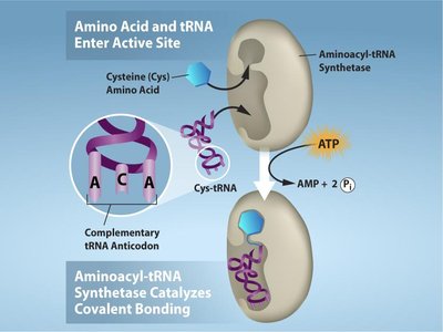
Aminoacyl-tRNA Synthetases
These enzymes catalyze the attachment of amino acids to their corresponding tRNAs, a process known as "charging" the tRNA. There are 20 different synthetases, one for each amino acid, and the reaction requires ATP.
Specificity: Each synthetase recognizes only one amino acid and its compatible tRNAs.
Reaction: Amino acid + tRNA + ATP → aminoacyl-tRNA + AMP + PPi
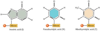
tRNA Modifications
tRNAs undergo extensive post-transcriptional modifications, including base modifications, trimming of precursor sequences, addition of the CCA tail, and removal of introns. These modifications enhance tRNA stability and accuracy during translation.
Stages of Translation
Initiation (Prokaryotes)
Translation initiation in prokaryotes involves the assembly of the ribosome on the mRNA at the start codon, facilitated by the Shine–Dalgarno sequence, which aligns the ribosome with the start codon. Initiation factors (IF1, IF2, IF3) assist in this process.
Initiator tRNA: Carries N-formylmethionine (fMet) in bacteria.
Shine–Dalgarno Sequence: Ribosome-binding site upstream of the start codon.
Elongation
During elongation, amino acids are sequentially added to the growing polypeptide chain. Each cycle involves the entry of a charged tRNA into the A site, peptide bond formation, and translocation of the ribosome.
Elongation Factors: EF-Tu (brings tRNA), EF-G (translocation).
Polysomes: Multiple ribosomes can translate a single mRNA simultaneously.
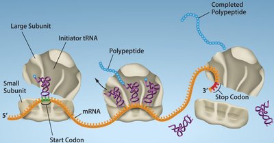
Termination
Termination occurs when a stop codon enters the A site. Release factors (RF1, RF2, RF3 in bacteria) recognize stop codons and promote the release of the completed polypeptide from the ribosome.
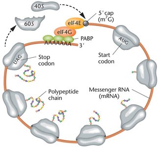
Translation in Eukaryotes
Eukaryotic translation is more complex, involving larger ribosomes, more initiation factors, and additional mRNA processing (5' cap, poly-A tail). The Kozak sequence helps identify the correct start codon.
Initiation Factors: eIF4E (cap binding), eIF4G (scaffold protein), PABP (poly-A binding protein).
Ribosome Recycling: Ribosomes may reinitiate translation on the same mRNA.
One-Gene–One-Enzyme and One-Gene–One-Polypeptide Hypotheses
Early genetic studies established that each gene encodes a specific enzyme (one-gene–one-enzyme hypothesis), later refined to one-gene–one-polypeptide as it became clear that not all proteins are enzymes and some proteins are composed of multiple polypeptides.
Protein Folding, Modification, and Targeting
Protein Folding
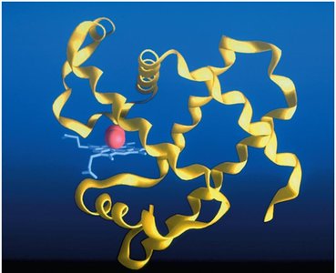
After translation, polypeptides fold into specific three-dimensional structures, which are essential for their function. Some proteins fold spontaneously, while others require chaperone proteins to assist in proper folding.
Primary Structure: Linear sequence of amino acids.
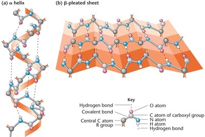
Secondary Structure: α-helix and β-pleated sheet, stabilized by hydrogen bonds.
Tertiary Structure: Overall three-dimensional shape.
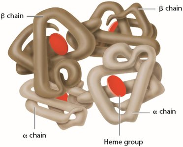
Quaternary Structure: Association of multiple polypeptide chains.
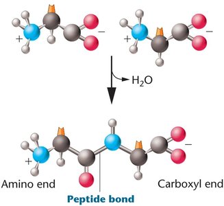
Peptide Bond Formation
A peptide bond forms between the carboxyl group of one amino acid and the amino group of another, releasing a molecule of water (condensation reaction).
Equation:
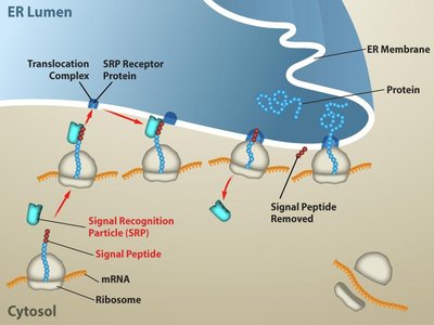
Protein Targeting
Proteins destined for secretion or specific organelles contain signal sequences that direct them to their proper cellular locations. The signal recognition particle (SRP) recognizes these sequences and targets the ribosome to the endoplasmic reticulum (ER) membrane in eukaryotes.
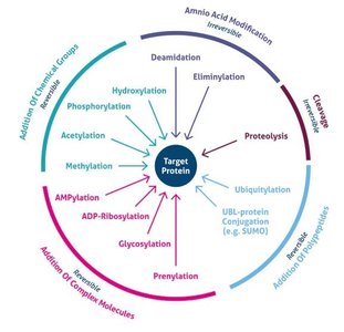
Post-Translational Modifications
Types and Functions
After translation, proteins often undergo post-translational modifications (PTMs) that are critical for their function, stability, localization, and regulation. Common PTMs include phosphorylation, acetylation, methylation, glycosylation, ubiquitination, SUMOylation, lipidation, and hydroxylation.
Modification | Main Function |
|---|---|
Phosphorylation | Regulates enzyme activity and cell signaling |
Acetylation | Controls gene expression and protein stability |
Methylation | Modulates protein interactions and gene regulation |
Glycosylation | Aids protein folding, stability, and cell recognition |
Ubiquitination | Tags proteins for degradation by the proteasome |
SUMOylation | Regulates protein localization and transcription |
Lipidation | Targets proteins to cell membranes |
Hydroxylation | Important for collagen stability and oxygen sensing |
Disulfide bonds | Stabilize protein structure |
Protein Domains and Functional Diversity
Protein Domains
Protein domains are distinct structural and functional units within a protein, typically consisting of 50–300 amino acids. Domains can confer specific functions such as DNA binding, dimerization, or interaction with other proteins or molecules.
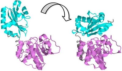
Protein-Protein Interactions
Protein-protein interactions (PPIs) are essential for many cellular processes, including signal transduction, gene regulation, and metabolic pathways. PPIs can induce conformational changes that activate or inhibit protein function.
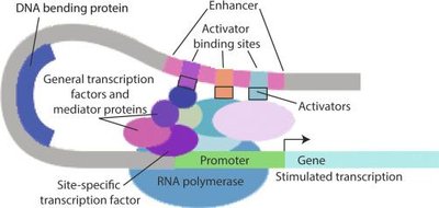
Regulation of Gene Expression by Protein Modifications
Ubiquitination and Protein Degradation
Ubiquitination is a post-translational modification where ubiquitin is attached to a protein, marking it for degradation by the proteasome. This process regulates protein levels and is a key mechanism for controlling gene expression.
Complex Regulation: NF-κB Pathway Example
The NF-κB signaling pathway illustrates how multiple post-translational modifications (ubiquitination, phosphorylation, cleavage, and translocation) can regulate gene expression. The stability and activity of key signaling proteins are tightly controlled by these modifications, affecting the transcription of target genes.
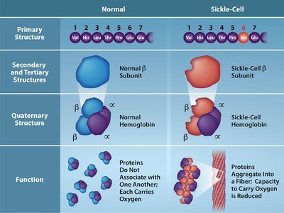
Practice Example: Sickle-Cell Anemia
Sickle-cell anemia is caused by a point mutation in the β-globin gene, resulting in the substitution of valine for glutamic acid at position 6. This single amino acid change alters the protein's structure and function, demonstrating the importance of translation accuracy and protein folding.
Summary Table: Key Differences in Translation
Feature | Prokaryotes | Eukaryotes |
|---|---|---|
Ribosome Size | 70S (50S + 30S) | 80S (60S + 40S) |
Initiation Site | Shine–Dalgarno sequence | Kozak sequence |
Initiator tRNA | fMet-tRNAfMet | Met-tRNAiMet |
Location | Cytoplasm | Transcription in nucleus, translation in cytoplasm |
mRNA Processing | None | 5' cap, poly-A tail, splicing |
Conclusion
Translation and post-translational modifications are central to the flow of genetic information from DNA to functional proteins. Understanding these processes is essential for comprehending gene expression, protein function, and the molecular basis of genetic diseases.
