 Back
BackCh 13 Translation and Proteins: From mRNA to Functional Polypeptides
Study Guide - Smart Notes
 Tailored notes based on your materials, expanded with key definitions, examples, and context.
Ch 13 Translation and Proteins
Introduction to Translation
The final product of gene expression is a polypeptide chain, which consists of a linear series of amino acids. The sequence of these amino acids is prescribed by the genetic code, and the process by which information in mRNA is used to synthesize polypeptides is called translation.
Translation is the biological polymerization of amino acids into polypeptide chains.
Key components required: ribosomes and transfer RNAs (tRNAs).
Transfer RNAs (tRNAs)
tRNAs serve as adaptors that match specific triplet codons in mRNA to their corresponding amino acids. Each tRNA contains an anticodon that base-pairs with the mRNA codon and carries the correct amino acid.
tRNAs are small, stable molecules (75–90 nucleotides).
Transcribed from DNA as large precursors and contain modified bases.
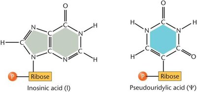
tRNAs fold into a characteristic cloverleaf structure with loops containing modified bases that do not form base pairs.
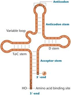
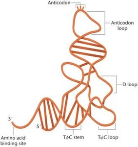
The three-dimensional structure of tRNA reveals an anticodon loop at one end and a 3′-acceptor region for amino acid attachment at the other.
Charging tRNA
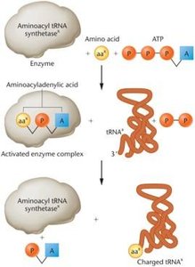
The process of attaching an amino acid to its corresponding tRNA is called charging and is catalyzed by aminoacyl tRNA synthetase enzymes. There are 20 different synthetases, one for each amino acid, and 30–55 different tRNAs.
Amino acid is activated by reacting with ATP to form aminoacyladenylic acid.
The enzyme then transfers the amino acid to the tRNA, forming a charged tRNA.
Ribosomes and rRNA
Ribosomal Structure
Ribosomes are essential for the expression of genetic information. They are composed of ribosomal proteins and ribosomal RNAs (rRNAs), and consist of large and small subunits.
Prokaryotic ribosomes: 70S (50S + 30S subunits)
Eukaryotic ribosomes: 80S (60S + 40S subunits)
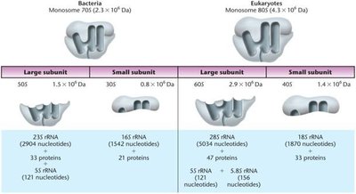
rRNA genes (rDNA) are present in clusters on chromosomes, with each cluster containing tandem repeats separated by noncoding spacer DNA.
Mechanism of Translation
Phases of Translation
Translation is divided into three main phases:
Initiation
Elongation
Termination
Each phase requires specific protein factors, as summarized in the table below.
Process | Factor | Role |
|---|---|---|
Initiation | IF1 | Binds to 30S subunit and prevents aminoacyl tRNA from binding to the A site prematurely |
Initiation | IF2 | Binds to initiator fMet-tRNA and transfers it to the P site; releases upon GTP hydrolysis |
Initiation | IF3 | Prevents premature association of 30S and 50S subunits |
Elongation | EF-Tu | Brings aminoacyl tRNA to the A site |
Elongation | EF-Ts | Regulates EF-Tu activity |
Elongation | EF-G | Stimulates translocation (GTP-dependent) |
Termination | RF1 | Releases polypeptide chain (UAA, UAG codons) |
Termination | RF2 | Releases polypeptide chain (UGA, UAA codons) |
Termination | RF3 | Stimulates RF1 and RF2 release |
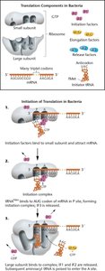
Initiation of Translation
Initiation requires both ribosomal subunits, mRNA, GTP, a charged initiator tRNA, and three initiation factors. In bacteria, the initiation codon (AUG) codes for formylmethionine (fMet). The Shine–Dalgarno sequence on mRNA base-pairs with the 16S rRNA to facilitate initiation.
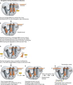
Elongation
During elongation, the polypeptide chain is lengthened one amino acid at a time. Charged tRNAs are brought to the A site by elongation factors, and peptide bonds are formed by peptidyl transferase activity of the 23S rRNA. The growing chain is transferred from the tRNA in the P site to the tRNA in the A site, and the uncharged tRNA exits via the E site.
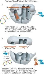
Termination
Termination occurs when a stop codon (UAG, UAA, UGA) enters the A site. Release factors stimulate the hydrolysis of the polypeptide from the tRNA, and the ribosomal subunits dissociate.
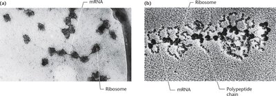
Polyribosomes (Polysomes)
Multiple ribosomes can translate a single mRNA simultaneously, forming polyribosomes or polysomes. This increases the efficiency of protein synthesis.
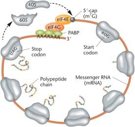
Eukaryotic Translation
Key Differences in Eukaryotes
Eukaryotic translation is more complex due to larger ribosomes, compartmentalization of transcription and translation, and additional mRNA processing (5′ cap and poly-A tail). The Kozak sequence (A/GNN AUG G) enhances translation initiation efficiency.
Poly-A tail and cap structure are essential for translation initiation and mRNA stability.
Closed-loop translation allows efficient ribosome recycling and prevents translation of degraded mRNA.
From Genes to Proteins
One-Gene:One-Enzyme and One-Gene:One-Polypeptide Hypotheses
Beadle and Tatum demonstrated that genes are directly responsible for the synthesis of enzymes, leading to the one-gene:one-enzyme hypothesis. This was later refined to the one-gene:one-polypeptide chain hypothesis as it became clear that not all proteins are enzymes and many proteins are composed of multiple polypeptide chains.
Polypeptides and Proteins
Polypeptides are precursors to proteins, assembled on ribosomes and folded into functional three-dimensional conformations to become proteins.
Amino Acids and Peptide Bonds
Amino acids contain a carboxyl group, an amino group, and a variable R group (side chain) that determines the amino acid's properties. R groups can be nonpolar (hydrophobic), polar (hydrophilic), or charged. Peptide bonds form between the amino group of one amino acid and the carboxyl group of another via a dehydration reaction.
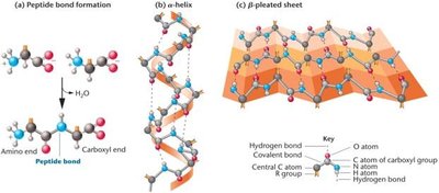
Levels of Protein Structure
Primary structure: Sequence of amino acids.
Secondary structure: Alpha helices and beta-pleated sheets.
Tertiary structure: Three-dimensional conformation.
Quaternary structure: Multiple polypeptide chains.
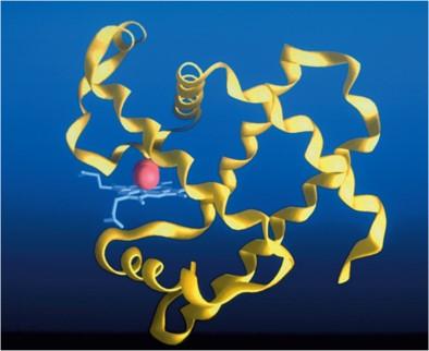
Protein Folding and Misfolding
Protein folding is mediated by chaperones. Misfolded proteins are tagged by ubiquitins and degraded in the proteasome. Misfolded proteins can cause diseases such as prion diseases (scrapie, mad cow disease, Creutzfeldt–Jakob disease) and neurodegenerative disorders (Huntington, Alzheimer, Parkinson).
Protein Function and Diversity
Functional Roles of Proteins
Hemoglobin and myoglobin: Oxygen transport.
Collagen and keratin: Structural proteins.
Actin and myosin: Muscle contraction.
Tubulin: Microtubule formation.
Immunoglobulins: Immune response.
Transport proteins: Molecule movement across membranes.
Hormones and receptors: Regulation of cellular activities.
Histones: DNA binding in eukaryotes.
Transcription factors: Regulation of gene expression.
Enzymes
Enzymes are proteins that catalyze chemical reactions, increasing reaction rates and determining the metabolic capacity of cells.
Protein Domains
Protein domains are sequences of 50–300 amino acids that fold into stable, unique conformations and impart specific functional capabilities, such as catalytic or DNA-binding domains.
