 Back
Translation and Proteins: Genetic Code, tRNA, Ribosomes, and Protein Structure
Study Guide - Smart Notes
 Tailored notes based on your materials, expanded with key definitions, examples, and context.
Translation and Proteins
The Central Dogma and Genetic Code
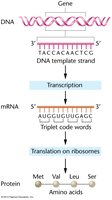
The central dogma of molecular biology describes the flow of genetic information from DNA to RNA to protein. Translation is the process by which messenger RNA (mRNA) is decoded to synthesize polypeptides, which fold into functional proteins. The genetic code is a set of rules by which information encoded in mRNA is translated into amino acids.
Genetic Code: Written in linear form using ribonucleotide bases (A, U, G, C) as "letters".
Triplet Codons: Each codon consists of three nucleotides and specifies one amino acid.
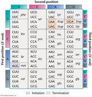
Start Codon: AUG (codes for methionine) initiates translation.
Stop Codons: UAA, UGA, UAG terminate translation and do not code for amino acids.
Degeneracy: Most amino acids are specified by more than one codon.
Example: The codon table shows all possible triplet combinations and their corresponding amino acids.
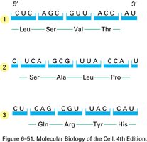
Reading Frame and Open Reading Frame (ORF)
The reading frame of mRNA is defined by the site of initiation (AUG). The open reading frame (ORF) is a sequence of codons that begins with a start codon and ends with a stop codon, determining the sequence of amino acids in the protein.
Transfer RNA (tRNA): Structure and Function
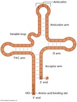
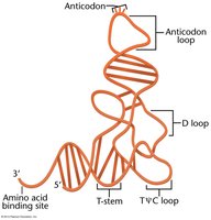
Transfer RNA (tRNA) acts as an adaptor molecule, translating the genetic code in mRNA into the corresponding amino acid sequence. tRNAs are transcribed from DNA and have a characteristic cloverleaf structure in 2-D and a complex 3-D shape.
Anticodon: Region of tRNA that base-pairs with the codon in mRNA.
Acceptor Arm: 3' end of tRNA where the amino acid is attached.
tRNA Charging: Aminoacyl tRNA synthetases catalyze the attachment of amino acids to tRNAs.
Number: At least 30 tRNA species accommodate 61 codons.
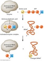
Charging of tRNA (Aminoacylation)
Before translation, tRNA molecules must be charged with their respective amino acids. This process is catalyzed by aminoacyl tRNA synthetases, which recognize both the tRNA and the amino acid.
Activation: Amino acid is activated by ATP.
Attachment: Activated amino acid is transferred to the tRNA.
Result: Charged tRNA can participate in translation.
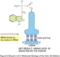
Codon-Anticodon Recognition
The anticodon of tRNA base-pairs with the complementary codon on mRNA, ensuring the correct amino acid is added to the growing polypeptide chain.
Degeneracy and the Wobble Hypothesis
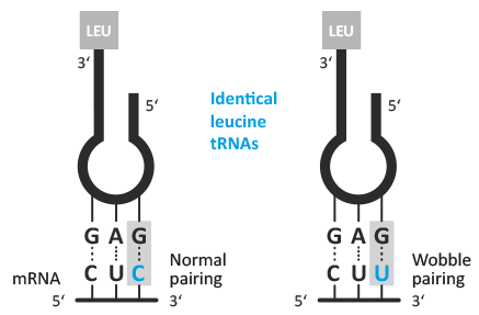
The genetic code is degenerate, meaning multiple codons can code for the same amino acid. The wobble hypothesis explains how the third position of the codon allows for flexible base pairing, enabling fewer tRNA species to recognize multiple codons.
Strict Pairing: First two positions follow Watson-Crick rules.
Wobble: Third position allows non-standard pairing.
Efficiency: At least 30 tRNAs accommodate 61 codons.
Ribosomal Structure and Function
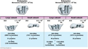
Ribosomes are the molecular machines responsible for protein synthesis. They consist of ribosomal RNA (rRNA) and proteins, forming large and small subunits. Ribosomes decode mRNA and catalyze peptide bond formation.
Prokaryotic Ribosomes: 70S (50S + 30S)
Eukaryotic Ribosomes: 80S (60S + 40S)
Svedberg Unit (S): Measures sedimentation rate during centrifugation.
Translation: Steps and Components
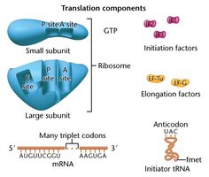
Translation is divided into three main steps: initiation, elongation, and termination. Each step requires specific factors and energy sources.
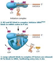
Initiation: Assembly of ribosome, mRNA, initiator tRNA, and initiation factors.
Elongation: Sequential addition of amino acids, catalyzed by peptidyl transferase.
Termination: Release of polypeptide upon encountering a stop codon.
Initiation in Prokaryotes
In prokaryotes, the initiation codon (AUG) codes for N-formylmethionine (fmet). The Shine-Dalgarno sequence helps position the ribosome for translation initiation.
Initiator tRNA: Carries fmet.
Shine-Dalgarno Sequence: AGGAGG upstream of AUG, recognized by the small ribosomal subunit.
Initiation in Eukaryotes
Eukaryotic mRNAs often contain a Kozak sequence, which enhances translation efficiency. Ribosomes are larger and more complex, and translation occurs in the cytoplasm.
Kozak Sequence: Purine-rich region upstream of AUG.
Ribosome Location: Free or bound to endoplasmic reticulum.
Elongation
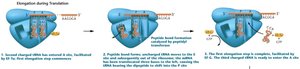
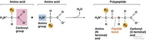
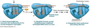
Elongation involves the addition of amino acids to the growing polypeptide chain. The ribosome has two main sites: the P (peptidyl) site and the A (aminoacyl) site. Peptidyl transferase catalyzes peptide bond formation.
Elongation Factors: EF-Tu, EF-G, etc.
Energy Source: GTP hydrolysis.
Termination
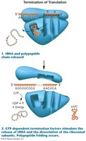
Termination occurs when a stop codon enters the A site. Release factors cleave the polypeptide from the tRNA, and the ribosome dissociates.
Stop Codons: UAG, UAA, UGA.
Release Factors: GTP-dependent proteins that facilitate polypeptide release.
Protein Structure: Primary, Secondary, Tertiary, and Quaternary
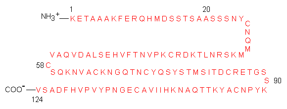
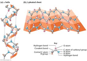
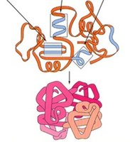
Proteins fold into specific structures that determine their function. The structure is organized into four levels:
Primary Structure: Linear sequence of amino acids, stabilized by peptide bonds.
Secondary Structure: Local folding into α-helices and β-sheets, stabilized by hydrogen bonds.
Tertiary Structure: Overall 3D shape, stabilized by covalent, ionic, disulfide, and hydrogen bonds.
Quaternary Structure: Association of multiple polypeptide chains.
Prokaryotic vs. Eukaryotic Translation
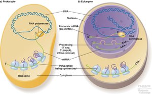
Translation differs between prokaryotes and eukaryotes in terms of ribosome structure, location, and complexity.
Prokaryotes: Transcription and translation occur simultaneously in the cytoplasm.
Eukaryotes: Transcription occurs in the nucleus; translation occurs in the cytoplasm.
Ribosomes: Eukaryotic ribosomes are larger and more complex.
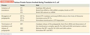
Protein Factors in Translation (E. coli)
Various protein factors are involved in the initiation, elongation, and termination of translation in prokaryotes.
Process | Factor | Role |
|---|---|---|
Initiation | IF1 | Stabilizes 30S subunit |
Initiation | IF2 | Binds fmet-tRNA to 30S-mRNA complex; binds to GTP |
Initiation | IF3 | Binds 30S subunit to mRNA |
Elongation | EF-Tu | Binds GTP, mediates aminoacyl-tRNA entry to A site |
Elongation | EF-G | Generates active EF-Tu; stimulates translocation, GTP-dependent |
Termination | RF1 | Catalyzes release of polypeptide chain from tRNA and dissociation of ribosome |
Termination | RF2 | Behaves like RF1; recognizes UAA and UGA termination codons |
Termination | RF3 | Stimulates RF1 and RF2 |
Review Questions
Describe the features of the genetic code.
What is tRNA? Describe the roles of tRNA as an adaptor molecule.
Describe the steps involved in charging a tRNA molecule with an amino acid.
What is the Wobble hypothesis? Explain how only 30 tRNA species can accommodate 61 triplet codons.
Compare and contrast prokaryotic and eukaryotic ribosomal structures.
Describe the steps involved in translation.
Describe the roles of translation protein factors involved in various steps.
How protein’s primary, secondary, and tertiary structures are defined?
Compare prokaryotic and eukaryotic translation processes.
