 Back
Translation and Proteins: Molecular Mechanisms and Genetic Implications
Study Guide - Smart Notes
 Tailored notes based on your materials, expanded with key definitions, examples, and context.
Translation and Proteins
Translation of mRNA Depends on Ribosomes and Transfer RNAs
Translation is the biological process by which amino acids are polymerized into polypeptide chains, forming proteins. This process is central to gene expression and requires several key components:
Amino acids: The building blocks of proteins.
Messenger RNA (mRNA): Carries genetic information from DNA to the ribosome.
Ribosomes: Complexes of ribosomal RNA (rRNA) and proteins that facilitate translation.
Transfer RNA (tRNA): Adaptor molecules that match mRNA codons to their corresponding amino acids.
Ribosomes: Structure and Function
Ribosomes play an essential role in the expression of genetic information. They consist of two subunits (large and small) and are composed of rRNA and proteins. The size and composition differ between prokaryotes and eukaryotes:
Prokaryotic ribosomes: 70S (50S large, 30S small)
Eukaryotic ribosomes: 80S (60S large, 40S small)
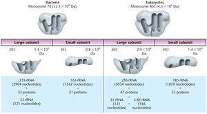
rDNA and rRNA Genes
rDNA refers to the genes encoding rRNA, which are present in clusters at various chromosomal sites. These clusters contain tandem repeats separated by noncoding spacer DNA.
tRNA: Structure and Function
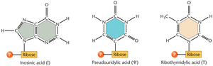
tRNAs are small, stable molecules (75–90 nucleotides) transcribed from DNA as larger precursors and then cleaved. They contain posttranscriptionally modified bases, such as inosine, pseudouridine, and ribothymidine.
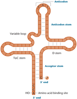
tRNAs have a characteristic cloverleaf structure, which includes the anticodon loop, D loop, TψC loop, and acceptor stem.
The three-dimensional structure of tRNA further stabilizes its function as an adaptor molecule.
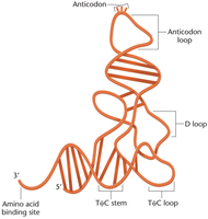
Anticodon and tRNA Charging
The anticodon region of tRNA base-pairs with the codon in mRNA, ensuring the correct amino acid is added to the growing polypeptide chain. The amino acid is covalently linked to the CCA sequence at the 3' end of all tRNAs.
Before translation, tRNAs must be "charged" with their respective amino acids by aminoacyl tRNA synthetases. There are 20 different synthetases, each highly specific for one amino acid.
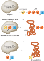
Translation of mRNA Can Be Divided into Three Steps
Translation occurs in three main stages: initiation, elongation, and termination.
Initiation of Translation
Initiation requires the assembly of the small and large ribosomal subunits, mRNA, GTP, a charged initiator tRNA, and initiation factors. In bacteria, the Shine–Dalgarno sequence precedes the AUG start codon and base-pairs with the 16S rRNA of the 30S subunit, facilitating initiation.
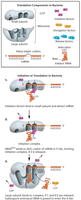
Elongation
During elongation, both ribosomal subunits are assembled with mRNA, forming the P site and A site. The 23S rRNA catalyzes peptide bond formation between the amino acid on tRNA at the A site and the growing peptide chain at the P site. Uncharged tRNA moves to the E (exit) site.
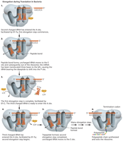
Termination
Termination is signaled by stop codons (UAG, UAA, UGA) in the A site. These codons do not specify any amino acid. GTP-dependent release factors stimulate hydrolysis of the polypeptide from the peptidyl tRNA, releasing it from the translation complex.
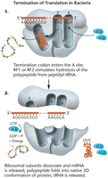
Polyribosomes (Polysomes)
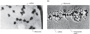
Polysomes are mRNAs with several ribosomes translating at once, increasing the efficiency of protein synthesis.
Translation Is More Complex in Eukaryotes
Eukaryotic translation involves larger, longer-lived ribosomes and additional factors. Transcription occurs in the nucleus, and mRNA is capped with a 7-methylguanosine residue and a poly-A tail, both essential for translation. The Kozak sequence increases the efficiency of translation initiation.
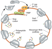
Closed loop translation occurs when the mRNA forms a loop, bringing the cap and tail together via poly-A-binding proteins and cap-binding proteins.
The Initial Insight That Proteins Are Important in Heredity: Inborn Errors of Metabolism
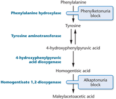
Mutations can lead to metabolic blocks, resulting in inborn errors of metabolism such as alkaptonuria and phenylketonuria (PKU). PKU is caused by inactive phenylalanine hydroxylase, preventing the conversion of phenylalanine to tyrosine and leading to intellectual disabilities.
Studies of Neurospora Led to the One-Gene: One-Enzyme Hypothesis
George Beadle and Edward Tatum demonstrated that genes are directly responsible for the synthesis of enzymes. Their experiments with Neurospora led to the one-gene:one-enzyme hypothesis, which was later refined to the one-gene:one-polypeptide hypothesis.
Studies of Human Hemoglobin Established That One Gene Encodes One Polypeptide
Research on human hemoglobin revealed that nearly all enzymes are proteins, but not all proteins are enzymes. Some proteins have subunit structures with two or more polypeptide chains. Sickle-cell anemia, inherited as a Mendelian trait, is caused by a single amino acid change in hemoglobin, demonstrating the genetic basis of protein structure and function.
Variation in Protein Structure Provides the Basis of Biological Diversity
Polypeptides are precursors to proteins, which fold into functional three-dimensional conformations. The structure of proteins is determined by the sequence and properties of amino acids:
Primary structure: Sequence of amino acids
Secondary structure: Local folding (e.g., alpha helices, beta sheets)
Tertiary structure: Three-dimensional conformation
Quaternary structure: Composed of more than one polypeptide chain
Posttranslational Modification Alters the Final Protein Product
Polypeptide chains are often modified after synthesis, which is crucial for the functional capability of the final protein product. Modifications include removal or modification of the N-terminus, attachment of carbohydrate side chains, trimming, removal of target peptides, and complexing with prosthetic groups. Protein folding may require chaperones, and misfolding can lead to diseases such as Creutzfeldt–Jakob disease and bovine spongiform encephalopathy (BSE).
Proteins Perform Many Diverse Roles
Proteins are the most abundant macromolecules in cells and perform diverse roles, including:
Transport: Hemoglobin and myoglobin transport oxygen.
Structure: Collagen and keratin provide structural support.
Movement: Actin and myosin are contractile proteins in muscle.
Cell division: Tubulin forms spindle fibers.
Immunity: Immunoglobulins function in the immune system.
Regulation: Hormones, receptors, histones, and transcription factors regulate cellular activities.
Enzymes: Catalyze chemical reactions, lowering activation energy.
Proteins Often Include More Than One Functional Domain
Protein domains are sequences of 50–300 amino acids that fold into stable, unique conformations, imparting different functional capabilities. The exon shuffling hypothesis suggests that exons from ancestral genes were recombined during evolution to create new protein domains.
Component | Function |
|---|---|
Ribosome | Protein synthesis |
tRNA | Adaptor for amino acid incorporation |
mRNA | Template for translation |
Aminoacyl tRNA synthetase | Charges tRNA with amino acid |
Initiation factors | Facilitate translation initiation |
Release factors | Terminate translation |
Additional info: The notes above expand on brief points with academic context, definitions, and examples to ensure completeness and clarity for Genetics students.
