 Back
BackTranslation of mRNA: Mechanisms and Genetic Implications
Study Guide - Smart Notes
 Tailored notes based on your materials, expanded with key definitions, examples, and context.
Translation of mRNA
Overview of Translation
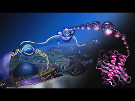
Translation is the process by which messenger RNA (mRNA) is decoded to synthesize proteins, a fundamental mechanism in gene expression. This process occurs in both prokaryotes and eukaryotes, with some differences in the machinery and regulation.
Translation involves the ribosome, tRNA, and various translation factors.
Proteins are synthesized by linking amino acids in the order specified by the mRNA sequence.
Translation occurs in three stages: initiation, elongation, and termination.
In prokaryotes, translation can begin before transcription is completed due to the absence of a nucleus.
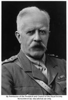
Archibald Garrod and the Relationship Between Genes and Proteins
Archibald Garrod was the first to propose a relationship between genes and protein production, based on his studies of patients with metabolic disorders such as alkaptonuria.
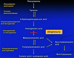
Alkaptonuria is a genetic disorder characterized by the accumulation of homogentisic acid due to a missing enzyme, homogentisic acid oxidase.
Garrod suggested that inherited traits are linked to defective enzymes, establishing the concept of "inborn errors of metabolism."
Clinical Manifestations of Alkaptonuria
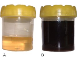
Darkening of urine upon standing due to homogentisic acid oxidation.
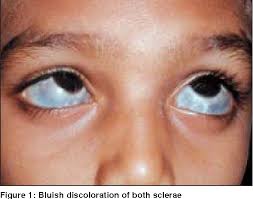
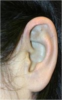
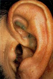
Blue-black discoloration of connective tissues (ochronosis).
Possible pigmentation of sclera and cartilage.

Gene Expression and Translation
Recognition Between tRNA and mRNA
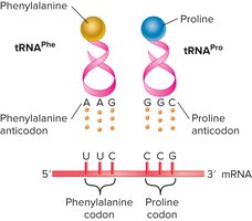
During translation, transfer RNA (tRNA) molecules recognize specific codons on the mRNA through their anticodon regions, ensuring the correct amino acid is incorporated into the growing polypeptide chain.
Anticodon: A sequence of three nucleotides in tRNA that pairs with the complementary codon in mRNA.
Each tRNA is named according to the amino acid it carries.
Structure of tRNA
tRNA molecules have a characteristic cloverleaf structure, with the anticodon region recognizing the mRNA codon and the amino acid attached at the 3’ end.
The anticodon loop binds to the mRNA codon.
The acceptor stem at the 3’ end attaches the amino acid.
Charging of tRNAs
The process of attaching an amino acid to its corresponding tRNA is called tRNA charging, catalyzed by aminoacyl-tRNA synthetases.
There are 20 aminoacyl-tRNA synthetases, one for each amino acid.
The reaction involves amino acid, tRNA, and ATP, resulting in a charged tRNA.
High accuracy is required; error rate is less than one in every 10,000.
Wobble Hypothesis and Codon-Anticodon Pairing
The genetic code is degenerate, meaning multiple codons can code for the same amino acid. Francis Crick's wobble hypothesis explains how tRNA can recognize more than one codon, especially at the third position.
The first two positions of the codon pair strictly, while the third position allows certain mismatches.
This flexibility increases the efficiency of translation.
Ribosome Structure and Functional Sites
Functional Sites of Ribosomes
Ribosomes are the molecular machines responsible for protein synthesis. They contain three discrete sites:
Peptidyl site (P site): Holds the tRNA carrying the growing polypeptide chain.
Aminoacyl site (A site): Holds the tRNA with the next amino acid to be added.
Exit site (E site): Where the uncharged tRNA exits the ribosome.
Initiation of Translation
Translation initiation involves the binding of mRNA to the ribosome. In prokaryotes, the Shine-Dalgarno sequence facilitates this binding, while in eukaryotes, the ribosome scans for the AUG start codon following Kozak’s rules.
Shine-Dalgarno sequence: Ribosomal binding site in prokaryotic mRNA, complementary to 16S rRNA.
Kozak’s rules: Define optimal context for translation initiation in eukaryotes.
Elongation Stage of Translation
During elongation, amino acids are added to the polypeptide chain one at a time. The ribosome moves along the mRNA, facilitating peptide bond formation and translocation of tRNAs.
Peptide bond formation is catalyzed by the 23S rRNA, making the ribosome a ribozyme.
Elongation rates: 15–20 amino acids/sec in bacteria; 2–6 amino acids/sec in eukaryotes.
Termination Stage of Translation
Translation terminates when a stop codon is reached. Stop codons are recognized by release factors, not tRNAs.
Three stop codons: UAG, UAA, UGA.
Bacteria have three release factors (RF1, RF2, RF3); eukaryotes have two (eRF1, eRF3).
Release factors mimic tRNA structure to facilitate termination.
Coupling of Transcription and Translation in Bacteria
In bacteria, transcription and translation are coupled because both processes occur in the cytoplasm. Translation can begin before transcription is completed, unlike in eukaryotes where transcription occurs in the nucleus and translation in the cytosol.
Antibiotics and Translation
Some antibiotics inhibit translation, thereby preventing the growth of microorganisms. These drugs target various components of the translation machinery.
Summary Table: Key Steps in Translation
Stage | Main Events | Key Molecules |
|---|---|---|
Initiation | mRNA binds ribosome; start codon recognized | Ribosome, mRNA, initiator tRNA |
Elongation | Amino acids added to polypeptide chain | Ribosome, tRNA, elongation factors |
Termination | Stop codon reached; polypeptide released | Release factors, ribosome, mRNA |
Key Equations
Peptide bond formation:
tRNA charging:
Additional info:
Clinical images (ears, sclera, urine, palms) illustrate the phenotypic effects of alkaptonuria, a classic example of gene-protein relationship.
tRNA-mRNA pairing diagram reinforces the concept of codon-anticodon recognition.
