 Back
BackTranslation, Post-Translational Modifications, and Protein Structure in Genetics
Study Guide - Smart Notes
 Tailored notes based on your materials, expanded with key definitions, examples, and context.
Translation: Decoding the Genetic Message
Overview of Translation
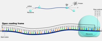
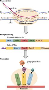
Translation is the process by which the genetic information encoded in messenger RNA (mRNA) is used to synthesize polypeptides (proteins). This process occurs in the cytoplasm and involves ribosomes, transfer RNAs (tRNAs), and various protein factors. The genetic code, composed of nucleotide triplets called codons, specifies the sequence of amino acids in a protein.
mRNA: Contains codons, each specifying an amino acid.
tRNA: Adapter molecules with anticodons complementary to mRNA codons; each carries a specific amino acid.
Ribosomes: Large RNA-protein complexes that facilitate mRNA binding and peptide bond formation.
The Genetic Code and Codon Recognition
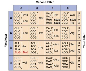
The genetic code is composed of 64 possible codons (43 combinations), which encode 20 amino acids and three stop signals. The code is degenerate, meaning that most amino acids are specified by more than one codon. The first and second bases of the codon are most important for determining the amino acid, while the third base is often less critical (the "wobble" position).
Stages of Translation
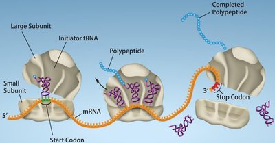
Translation proceeds in three main stages: initiation, elongation, and termination. Each stage involves specific molecular events and factors.
Initiation: Assembly of the ribosome on the mRNA at the start codon (AUG).
Elongation: Sequential addition of amino acids to the growing polypeptide chain.
Termination: Release of the completed polypeptide when a stop codon is encountered.
Translation Machinery: Ribosomes and tRNAs
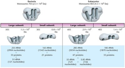
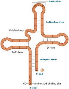
Ribosomes are composed of large and small subunits, each containing ribosomal RNA (rRNA) and proteins. Prokaryotic ribosomes are 70S (50S + 30S), while eukaryotic ribosomes are 80S (60S + 40S). tRNAs are small, stable RNA molecules (75–90 nucleotides) with a cloverleaf structure, containing an anticodon loop and an acceptor stem for amino acid attachment.
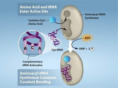
Charging tRNAs: Aminoacyl-tRNA Synthetases
Aminoacyl-tRNA synthetases are enzymes that catalyze the attachment of amino acids to their corresponding tRNAs, a process requiring ATP. There are 20 different synthetases, one for each amino acid, ensuring high specificity.
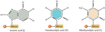
tRNA Modifications and Processing
tRNAs undergo extensive post-transcriptional modifications, including base modifications, trimming of precursor sequences, addition of the CCA tail at the 3' end, and removal of introns. These modifications enhance tRNA stability, translation accuracy, and codon recognition flexibility.
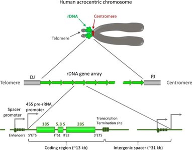
Ribosomal DNA (rDNA) Organization
rDNA refers to the DNA sequences encoding rRNA molecules. These genes are organized as tandem repeats in clusters, often located in nucleolus organizer regions (NORs) of chromosomes. Each repeat contains coding regions for rRNAs and noncoding spacers that regulate transcription.
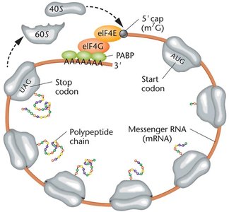
Translation Initiation in Prokaryotes and Eukaryotes
In prokaryotes, the Shine–Dalgarno sequence helps position the ribosome at the start codon. Initiation factors (IF1, IF2, IF3) assist in complex formation. In eukaryotes, the 5' cap and Kozak sequence facilitate ribosome binding, and additional initiation factors are required. The poly-A tail also plays a role in translation initiation.
Translation Elongation and Termination
During elongation, charged tRNAs enter the ribosome, peptide bonds are formed, and the ribosome translocates along the mRNA. Termination occurs when a stop codon is reached, and release factors promote the release of the polypeptide.
Protein Folding, Modification, and Targeting
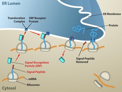
Protein Folding and Signal Sequences
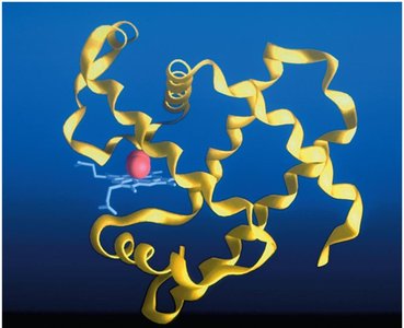
After translation, proteins fold into specific three-dimensional structures, sometimes with the help of chaperone proteins. Signal sequences direct proteins to specific cellular compartments, such as the endoplasmic reticulum (ER) for secretion. The signal recognition particle (SRP) recognizes these sequences and targets the ribosome to the ER membrane.
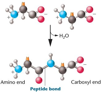
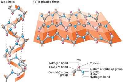
Peptide Bond Formation and Protein Structure Levels
Peptide bonds are formed by dehydration reactions between the carboxyl group of one amino acid and the amino group of another. Proteins have four levels of structure: primary (amino acid sequence), secondary (α-helix and β-pleated sheet), tertiary (three-dimensional conformation), and quaternary (multiple polypeptide chains).
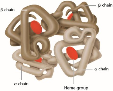
Genetic Mutations and Protein Function: Sickle-Cell Anemia
Point mutations can dramatically affect protein function. For example, sickle-cell anemia is caused by a single nucleotide change in the β-globin gene, resulting in the substitution of valine for glutamic acid at position 6. This alters hemoglobin structure and function, leading to disease symptoms.

Post-Translational Modifications (PTMs)
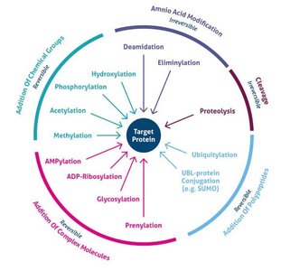
Types and Functions of PTMs
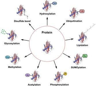
After translation, polypeptides often undergo post-translational modifications that are crucial for their final function. These modifications include phosphorylation, acetylation, methylation, glycosylation, ubiquitination, SUMOylation, lipidation, hydroxylation, and disulfide bond formation. Each modification can regulate protein activity, stability, localization, or interactions.
Modification | Main Function |
|---|---|
Phosphorylation | Regulates enzyme activity and cell signaling |
Acetylation | Controls gene expression and protein stability |
Methylation | Modulates protein interactions and gene regulation |
Glycosylation | Aids protein folding, stability, and cell recognition |
Ubiquitination | Tags proteins for degradation by the proteasome |
SUMOylation | Regulates protein localization and transcription |
Lipidation | Targets proteins to cell membranes |
Hydroxylation | Important for collagen stability and oxygen sensing |
Disulfide bonds | Stabilize protein structure |
Protein Domains and Functional Diversity
Protein Domains
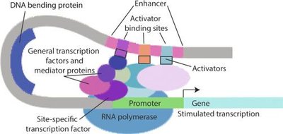
Protein domains are distinct structural and functional units within a protein, typically 50–300 amino acids in length. Domains can confer specific functions such as DNA binding, dimerization, ligand binding, or protein-protein interactions. Transcription factors, for example, are modular proteins with DNA-binding and effector domains.
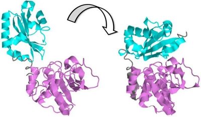
Protein-Protein Interactions (PPI)
PPIs are essential for many cellular processes, including signal transduction, enzyme activation, and transport. Structural changes induced by PPIs can activate or inhibit protein function, as seen in hemoglobin and protein kinases.
Regulation of Gene Expression and Protein Degradation
Gene Expression Regulation
Gene expression is regulated at multiple levels, including chromatin structure, transcription, mRNA processing, translation, and protein stability. The final protein level in a cell depends on both synthesis and degradation rates.
Ubiquitination and Proteasomal Degradation
Ubiquitination is a post-translational modification that tags proteins for degradation by the proteasome. Polyubiquitination (especially K48-linked) signals for proteasomal degradation, while other forms can regulate signaling without degradation. This process is a key mechanism for controlling protein levels and gene expression.
Example: NF-κB Pathway Regulation
The noncanonical NF-κB pathway illustrates regulation through ubiquitination, phosphorylation, cleavage, and translocation. In the absence of signaling, NIK is ubiquitinated and degraded, keeping the pathway off. Upon stimulation, NIK accumulates, activating downstream kinases and transcription factors, leading to gene expression changes.
Summary: Translation and post-translational modifications are central to gene expression and protein function. Understanding these processes is essential for interpreting genetic information and its impact on cellular physiology and disease.
