 Back
BackTranslation: The Molecular Biology of Protein Synthesis
Study Guide - Smart Notes
 Tailored notes based on your materials, expanded with key definitions, examples, and context.
Translation: The Molecular Biology of Protein Synthesis
Overview of Gene Expression
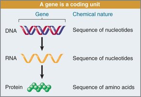
Gene expression is the process by which genetic information encoded in DNA is used to produce functional proteins. This occurs through two main steps: transcription (DNA to RNA) and translation (RNA to protein). The central dogma of molecular biology describes this flow of information: DNA → RNA → Protein.
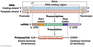
Transcription: Synthesis of RNA from a DNA template.
Translation: Synthesis of a polypeptide (protein) from an mRNA template.
The Structure and Properties of Amino Acids
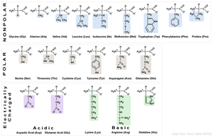
Proteins are composed of 20 common amino acids, each with unique chemical properties. Amino acids are classified based on their side chains as nonpolar, polar, acidic, or basic.
Nonpolar: Glycine, Alanine, Valine, Leucine, Isoleucine, Methionine, Tryptophan, Phenylalanine, Proline
Polar: Serine, Threonine, Cysteine, Tyrosine, Asparagine, Glutamine
Acidic: Aspartic acid, Glutamic acid
Basic: Lysine, Arginine, Histidine
Polypeptide Chain Formation
Amino acids are joined by peptide bonds, forming linear polypeptide chains. Each chain has an amino (N) terminus and a carboxyl (C) terminus, defining the directionality of the protein.
Peptide bond: Covalent bond between the carboxyl group of one amino acid and the amino group of the next.
Directionality: Proteins are synthesized from N-terminus to C-terminus.
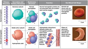
Protein Structure and Function
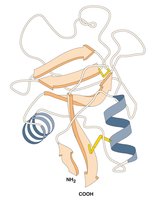
The three-dimensional structure of a protein determines its function. Proteins fold based on the chemical properties of their amino acids, and the forces stabilizing their conformation are weaker than covalent bonds.
Primary structure: Sequence of amino acids.
Secondary/tertiary/quaternary structure: Higher-order folding and assembly.
Function: Enzymatic activity, structural support, signaling, etc.
Genetic Mutations and Disease: Sickle Cell Anemia
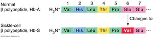
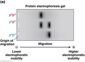
A single nucleotide change in DNA can alter the amino acid sequence of a protein, potentially causing disease. Sickle cell anemia is caused by a single amino acid substitution (Glu to Val) in the β-globin protein, affecting its structure and function.
Mutation: Point mutation in the β-globin gene.
Effect: Alters hemoglobin properties, leading to sickle-shaped red blood cells.
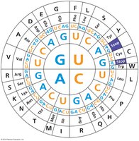
The Genetic Code
Triplet Nature of the Genetic Code
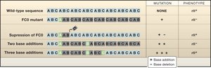
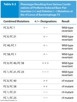
The genetic code is read in groups of three bases (codons), each specifying one amino acid. Experiments by Crick and Brenner using bacteriophage T4 mutants demonstrated the triplet nature of the code.
Codon: Three-base sequence in mRNA specifying an amino acid.
Triplet code: 43 = 64 possible codons, sufficient for 20 amino acids.


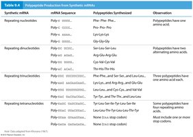
Deciphering the Genetic Code
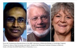
Marshall Nirenberg and Gobind Khorana used synthetic RNAs and cell-free extracts to determine which codons specify which amino acids. Their experiments established the complete genetic code.
PolyU experiment: UUU codes for phenylalanine.
PolyA experiment: AAA codes for lysine.
PolyC experiment: CCC codes for proline.
Repeating sequences: Used to identify codons for other amino acids.
Properties of the Genetic Code
The genetic code has several key properties:
Triplet: Each codon is three bases.
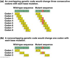
Non-overlapping: Codons are read one after another, not overlapping.
No punctuation: The code is read continuously.
Start and stop codons: AUG (start), UAG/UAA/UGA (stop).
Degenerate: Most amino acids are encoded by more than one codon.
Nearly universal: The code is conserved across most organisms.
Translation: Mechanism and Machinery
Ribosome Structure and Function
The ribosome is the molecular machine responsible for translation. It reads the mRNA sequence and assembles the corresponding amino acid chain.
Ribosome: Composed of rRNA and proteins, with large and small subunits.
Active sites: A (aminoacyl), P (peptidyl), and E (exit) sites for tRNA binding.

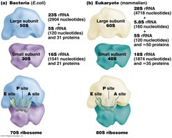
Translation in Prokaryotes vs Eukaryotes
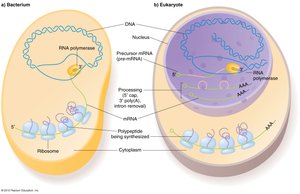
Translation occurs differently in prokaryotes and eukaryotes. In prokaryotes, transcription and translation are coupled, while in eukaryotes, they are separated by the nuclear membrane.
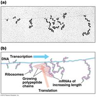
Prokaryotes: Translation begins before transcription is complete.
Eukaryotes: mRNA is processed in the nucleus, then translated in the cytoplasm.

Types of RNA in Translation
Several types of RNA are involved in translation:
mRNA: Messenger RNA, carries the genetic code.
tRNA: Transfer RNA, brings amino acids to the ribosome.
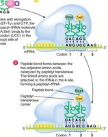
rRNA: Ribosomal RNA, forms the core of the ribosome and catalyzes peptide bond formation.
miRNA/siRNA: Regulate gene expression.
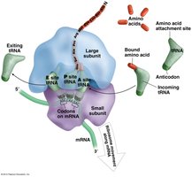
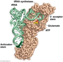
tRNA Structure and Function
tRNAs are adapter molecules that recognize specific codons in mRNA and deliver the corresponding amino acid. Each tRNA has an anticodon loop and an amino acid attachment site.
Anticodon: Three-base sequence complementary to the mRNA codon.
Charging: Aminoacyl-tRNA synthetase enzymes attach the correct amino acid to the tRNA.
Wobble Hypothesis
The third base of the codon (wobble position) allows for flexible pairing, enabling one tRNA to recognize multiple codons for the same amino acid.
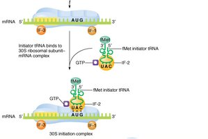
Stages of Translation
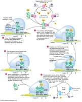
Translation occurs in three stages: initiation, elongation, and termination.
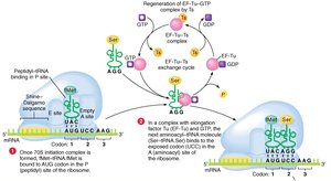
Initiation: Ribosome assembles at the start codon.
Elongation: Amino acids are added one by one to the growing polypeptide chain.
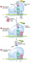
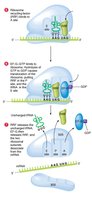
Termination: Translation ends at a stop codon, and the polypeptide is released.


Summary Table: Properties of the Genetic Code
Property | Description |
|---|---|
Triplet | Each codon is three bases |
Non-overlapping | Codons are read one after another |
No punctuation | Code is read continuously |
Start/Stop codons | AUG (start), UAG/UAA/UGA (stop) |
Degenerate | Most amino acids have multiple codons |
Universal | Code is conserved across organisms |
Key Equations
Number of possible codons:
Peptide bond formation:
Example: Sickle Cell Anemia
A single nucleotide change in the β-globin gene leads to a single amino acid substitution, which alters the protein's structure and function, causing sickle cell disease.
Additional info:
Some details about the triplet code and the experiments by Crick, Brenner, Nirenberg, and Khorana were inferred and expanded for academic completeness.
