Ch. 19 Blood
Martini - Fundamentals of Anatomy & Physiology 11th Edition
Martini, Nath, Bartholomew11th EditionFundamentals of Anatomy & PhysiologyISBN: 9780136874089당신이 사용하는 게 아니라요?교과서 변경
19장, 문제 1

Identify the five types of white blood cells in the following photographs:
<IMAGE>
a. ___
b. ___
c. ___
d. ___
e. ___

검증된 단계별 안내
1
Step 1: Understand the five main types of white blood cells (WBCs), also known as leukocytes. These are neutrophils, lymphocytes, monocytes, eosinophils, and basophils. Each has distinct morphological features visible under a microscope.
Step 2: Examine the characteristics of each WBC type in the photographs. For example, neutrophils typically have a multi-lobed nucleus and fine granules; lymphocytes have a large, round nucleus with little cytoplasm; monocytes are larger with a kidney-shaped nucleus; eosinophils have bi-lobed nuclei and large red-orange granules; basophils have large dark blue or purple granules that often obscure the nucleus.
Step 3: Match each photograph (a, b, c, d, e) to the WBC type by comparing the visible features in the images to the known characteristics of each cell type.
Step 4: Label each photograph accordingly with the identified WBC type: neutrophil, lymphocyte, monocyte, eosinophil, or basophil.
Step 5: Review your identifications by cross-checking the morphological features and ensuring that each WBC type is assigned to only one photograph, as there are five types and five images.

비슷한 문제에 대한 검증된 영상 답변:

이 영상 해법은 위 문제에 도움이 된다고 튜터들이 추천한 것입니다.

주요 개념

질문에 올바르게 답하기 위해 반드시 이해해야 하는 핵심 개념들은 다음과 같습니다.

Types of White Blood Cells (Leukocytes)

White blood cells are immune cells that protect the body against infections. The five main types are neutrophils, lymphocytes, monocytes, eosinophils, and basophils, each with distinct functions and appearances. Recognizing these types is essential for identifying them in microscopic images.
추천 영상:
가이드 코스
5:34
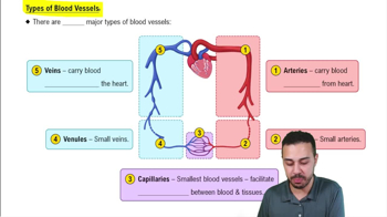
Types of Blood Vessels

Morphological Characteristics of White Blood Cells

Each white blood cell type has unique features such as nucleus shape, cytoplasm color, and granule presence. For example, neutrophils have multi-lobed nuclei, eosinophils contain red-orange granules, and lymphocytes have large, round nuclei. Understanding these traits aids in accurate identification.
추천 영상:
02:19
Characteristic 4: Supported by Connective Tissue

Staining Techniques in Hematology

Blood smears are stained using dyes like Wright or Giemsa stain to highlight cell structures. These stains color different cell components distinctly, making it easier to differentiate white blood cell types under a microscope. Familiarity with staining effects is crucial for interpreting images.
추천 영상:
가이드 코스
04:10
Epidermal Layers: Stratum Granulosum (Granular Layer)