Ch. 20 The Heart
Martini - Fundamentals of Anatomy & Physiology 11th Edition
Martini, Nath, Bartholomew11th EditionFundamentals of Anatomy & PhysiologyISBN: 9780136874089당신이 사용하는 게 아니라요?교과서 변경
20장, 문제 20

If the papillary muscles fail to contract:
(a) The ventricles will not pump blood
(b) The atria will not pump blood
(c) The semilunar valves will not open
(d) The AV valves will not close properly
(e) None of these happen

검증된 단계별 안내
1
Step 1: Understand the role of papillary muscles in the heart. Papillary muscles are located in the ventricles and attach to the atrioventricular (AV) valves (tricuspid and mitral valves) via chordae tendineae.
Step 2: Recognize that during ventricular contraction, papillary muscles contract to pull on the chordae tendineae, which prevents the AV valves from inverting or prolapsing back into the atria.
Step 3: If the papillary muscles fail to contract, the tension on the chordae tendineae is lost, which means the AV valves may not close properly and could allow blood to flow backward into the atria (a condition called valve regurgitation).
Step 4: Note that the ventricles can still pump blood even if the papillary muscles fail, but the efficiency is reduced due to valve leakage; the atria and semilunar valves are not directly affected by papillary muscle contraction.
Step 5: Conclude that the correct consequence of papillary muscle failure is that the AV valves will not close properly, corresponding to option (d).

비슷한 문제에 대한 검증된 영상 답변:

이 영상 해법은 위 문제에 도움이 된다고 튜터들이 추천한 것입니다.
도움이 되었나요?

주요 개념

질문에 올바르게 답하기 위해 반드시 이해해야 하는 핵심 개념들은 다음과 같습니다.

Function of Papillary Muscles

Papillary muscles are located in the ventricles of the heart and attach to the atrioventricular (AV) valves via chordae tendineae. Their contraction prevents the AV valves from prolapsing or inverting into the atria during ventricular contraction, ensuring proper valve closure and preventing backflow of blood.
추천 영상:
가이드 코스
9:42
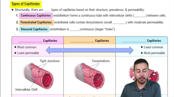
Types of Capillaries

Role of Atrioventricular (AV) Valves

The AV valves (tricuspid and mitral valves) regulate blood flow between the atria and ventricles. They open to allow blood to flow into the ventricles and close during ventricular contraction to prevent blood from flowing back into the atria, maintaining unidirectional blood flow.
추천 영상:
7:13
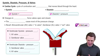
Systole, Diastole, Pressure, & Valves

Mechanism of Ventricular Contraction and Valve Operation

During ventricular systole, the ventricles contract to pump blood into the arteries. The papillary muscles contract simultaneously to hold the AV valves closed, while the semilunar valves open to allow blood to exit the heart. Failure of papillary muscles affects AV valve closure but does not stop ventricular contraction or semilunar valve function.
추천 영상:
4:27
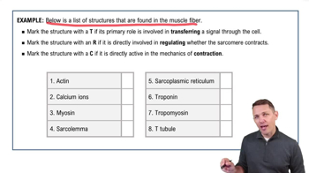
Steps of Muscle Contraction Example 1