Ch. 20 The Heart
Martini - Fundamentals of Anatomy & Physiology 11th Edition
Martini, Nath, Bartholomew11th EditionFundamentals of Anatomy & PhysiologyISBN: 9780136874089당신이 사용하는 게 아니라요?교과서 변경
20장, 문제 4

The simple squamous epithelium covering the heart valves is the:
(a) Epicardium
(b) Endocardium
(c) Myocardium
(d) Endothelium

검증된 단계별 안내
1
Step 1: Understand the structure of the heart wall, which consists of three layers: the epicardium (outer layer), myocardium (middle muscular layer), and endocardium (inner layer).
Step 2: Recognize that the heart valves are covered by a specialized type of simple squamous epithelium that lines the inner surfaces of the heart, including the valves.
Step 3: Recall that the endocardium is the inner lining of the heart chambers and valves, composed of simple squamous epithelium known as endothelium.
Step 4: Differentiate between the terms: 'endocardium' refers to the entire inner lining, while 'endothelium' specifically describes the simple squamous epithelial cells forming that lining.
Step 5: Conclude that the simple squamous epithelium covering the heart valves is called the endothelium, which is part of the endocardium.

비슷한 문제에 대한 검증된 영상 답변:

이 영상 해법은 위 문제에 도움이 된다고 튜터들이 추천한 것입니다.
도움이 되었나요?

주요 개념

질문에 올바르게 답하기 위해 반드시 이해해야 하는 핵심 개념들은 다음과 같습니다.

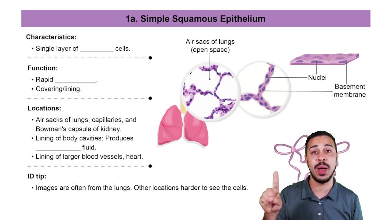
Simple Squamous Epithelium

Simple squamous epithelium is a single layer of flat, thin cells that allows for easy diffusion and smooth surfaces. It lines structures where rapid exchange or minimal friction is needed, such as blood vessels and heart valves.
추천 영상:
10:23
Simple Squamous Epithelia

Endocardium

The endocardium is the innermost layer of the heart, composed mainly of simple squamous epithelium called endothelium. It lines the heart chambers and covers the heart valves, providing a smooth surface for blood flow.
추천 영상:
가이드 코스
14:36
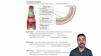
General Blood Vessel Structure

Heart Wall Layers

The heart wall consists of three layers: epicardium (outer layer), myocardium (muscle layer), and endocardium (inner layer). Understanding these layers helps identify which covers the valves and their specific tissue types.
추천 영상:
가이드 코스
02:31
Epidermal Layers: Stratum Lucidum (Clear Layer)
관련 실천
교과서 질문

The heart is surrounded by the:

(a) Pleural cavity

(b) Peritoneal cavity

(c) Abdominopelvic cavity

(d) Mediastinum

(e) Abdominal cavity

549
views
교과서 질문

The serous membrane covering the outer surface of the heart is the:

(a) Parietal layer of the serous pericardium

(b) Endocardium

(c) Myocardium

(d) Visceral layer of the serous pericardium

1539
views
1
rank
교과서 질문

The autonomic centers for cardiac function are located in:

(a) The myocardial tissue of the heart

(b) The cardiac centers of the medulla oblongata

(c) The cerebral cortex

(d) All of these structures

985
views
교과서 질문

Cardiac output is equal to the:

(a) Difference between the end-diastolic volume and the end-systolic volume

(b) Product of heart rate and stroke volume

(c) Difference between the stroke volume at rest and the stroke volume during exercise

(d) Stroke volume less the end-systolic volume

(e) Product of heart rate and blood pressure

1164
views
교과서 질문

TThe great cardiac vein drains blood from the heart muscle to the:

(a) Left ventricle

(b) Right ventricle

(c) Right atrium

(d) Left atrium

927
views
교과서 질문

The cardiac skeleton of the heart has which two of the following functions?

(a) It physically isolates the muscle fibers of the atria from those of the ventricles.

(b) It maintains the normal shape of the heart.

(c) It helps distribute the forces of cardiac contraction.

(d) It allows more rapid contraction of the ventricles.

(e) It strengthens and helps prevent overexpansion of the heart.

1010
views