 Back
BackCell Cycle, Mitosis, and Meiosis: Foundations of Genetic Transmission
Study Guide - Smart Notes
 Tailored notes based on your materials, expanded with key definitions, examples, and context.
Chapter 2: Fun with Mitosis, Meiosis, and the Cell Cycle
Introduction to Cell Division and Genetic Transmission
The transmission of genetic material in eukaryotes from one generation of cells to the next is accomplished through mitosis and meiosis. Mitosis produces two genetically identical cells, while meiosis generates gametes with half the chromosome number, enabling sexual reproduction and genetic diversity.
Cell Structure and Genetic Function
Types of Cells and Common Features
Cell structure is intimately linked to genetic function. There are two main cell types:
Prokaryotic cells (bacteria, archaea): Lack a nucleus and membrane-bound organelles.
Eukaryotic cells (protists, plants, fungi, animals): Possess a nucleus and various organelles.
All cells share fundamental features:
Plasma membrane
DNA
Ribosomes
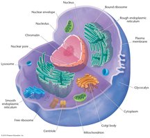
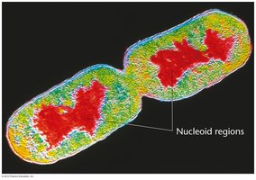
Chromatin and Chromosomes
In eukaryotes, DNA is complexed with proteins to form chromatin. During cell division, chromatin condenses into visible chromosomes. Chromosomes are the vehicles of genetic information.
Centrioles, Centrosomes, and Chromosome Movement
Animal cells contain centrioles within the centrosome, which organize spindle fibers for chromosome movement during mitosis and meiosis. Key terms to distinguish:
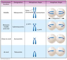
Centromere: Region where sister chromatids are joined.
Chromatid: One of two identical halves of a chromosome.
Chromosome: Structure carrying genetic material.
Sister chromatid: Two identical chromatids joined at the centromere.
Homologous chromosome: Chromosomes carrying genes for the same traits.
Chromosomes in Diploid Organisms
Homologous Pairs and Chromosome Number
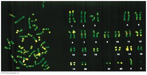
Somatic cells in diploid organisms contain chromosomes in homologous pairs. For example, humans have 46 chromosomes (23 pairs). Homologous chromosomes carry genes for the same inherited characteristics but may have different versions, called alleles.
Chromosome Numbers Across Species
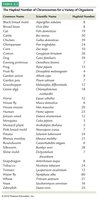
The haploid number of chromosomes varies among species. The table below summarizes chromosome numbers for various organisms:
Common Name | Scientific Name | Haploid Number |
|---|---|---|
Human | Homo sapiens | 23 |
Dog | Canis familiaris | 39 |
Chicken | Gallus domesticus | 39 |
Fruit fly | Drosophila melanogaster | 4 |
Mouse | Mus musculus | 20 |
Corn | Zea mays | 10 |
Frog | Rana pipiens | 13 |
Onion | Allium cepa | 8 |
Chimpanzee | Pan troglodytes | 24 |
Horse | Equus caballus | 32 |
Cat | Felis domestica | 19 |
Rabbit | Oryctolagus cuniculus | 22 |
Sheep | Ovis aries | 27 |
Goat | Capra hircus | 30 |
Guinea pig | Cavia porcellus | 30 |
Additional info: Table truncated for brevity. |
Mitosis: Partitioning Chromosomes
splits sister chromatids
The Cell Cycle
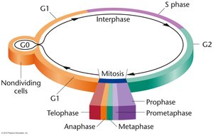
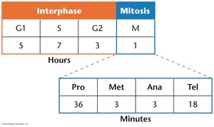
The cell cycle consists of interphase and mitosis. Interphase includes:
G1 phase: Cell growth
S phase: DNA synthesis
G2 phase: Preparation for mitosis
G0 phase: Nondividing, metabolically active state
Stages of Mitosis
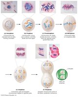
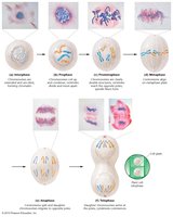
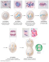
Mitosis is divided into discrete stages:
Prophase: Chromosomes condense, centrioles divide, nuclear envelope breaks down.
Prometaphase: Chromosomes move to the equatorial plane; nuclear envelope is gone.
Metaphase: Chromosomes align at the equatorial plane; spindle fibers attach to kinetochores.
Anaphase: Sister chromatids separate and migrate to opposite poles.
Telophase: Chromosomes uncoil, nuclear envelope reforms, cytokinesis occurs.
Mechanism of Chromatid Separation
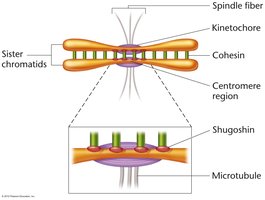
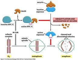
During metaphase, sister chromatids are held together by cohesin proteins. The enzyme separase cleaves cohesin, allowing chromatids to separate in anaphase. Shugoshin protects cohesin at the centromere until anaphase.
Meiosis: Reduction Division and Genetic Variation
genetic variability
Overview of Meiosis
Meiosis reduces the chromosome number from diploid (2n) to haploid (n), producing gametes or spores. It consists of two successive divisions: meiosis I and meiosis II.
Prophase I Substages
Prophase I is subdivided into:
Leptonema: Chromosomes appear as long, single threads.
Zygonema: Homologous chromosomes pair (synapsis), forming bivalents.
Pachynema: Chromosomes thicken, split into sister chromatids (tetrads); crossing over occurs.
Diplonema: Homologs begin to separate; chiasmata (sites of crossing over) become visible.
Diakinesis: Homologs further separate; nuclear envelope breaks down; tetrads align at equator.
Crossing over involves the exchange of genetic material between non-sister chromatids, increasing genetic diversity.
Meiosis I and II
Meiosis I separates homologous chromosomes, resulting in two haploid cells. Meiosis II separates sister chromatids, producing four haploid gametes.
Development of Gametes: Spermatogenesis and Oogenesis
Spermatogenesis
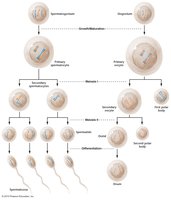
The primary spermatocyte undergoes meiosis I to produce two secondary spermatocytes, which undergo meiosis II to yield four haploid spermatids.
Oogenesis
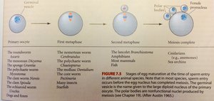
Oogenesis produces four daughter cells, but only one receives most of the cytoplasm and becomes the ovum. The other three, called polar bodies, do not undergo further division.
Meiosis and Sexual Reproduction
miosis 1 splitting homologous chromosomes
Genetic Variation and Life Cycles
Meiosis is essential for sexual reproduction in diploid organisms, generating genetic variation. Gametes receive either maternal or paternal chromosomes, and the number of possible combinations is , where n is the haploid number.
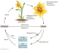
In multicellular plants, the life cycle alternates between diploid sporophyte and haploid gametophyte stages. Meiosis and fertilization bridge these stages.
Summary Table: Key Differences Between Mitosis and Meiosis
Feature | Mitosis | Meiosis |
|---|---|---|
Number of divisions | 1 | 2 |
Number of daughter cells | 2 | 4 |
Chromosome number in daughter cells | Diploid (2n) | Haploid (n) |
Genetic identity | Identical to parent | Genetically unique |
Function | Growth, repair | Sexual reproduction |
Additional info: Academic context was added to clarify the stages of cell division, the mechanism of chromatid separation, and the significance of meiosis in generating genetic diversity.
