 Back
BackLecture 3
Study Guide - Smart Notes
 Tailored notes based on your materials, expanded with key definitions, examples, and context.
The Chromosome Theory of Inheritance
Historical Development and Physical Basis
The chromosome theory of inheritance connects Mendel’s laws to the behavior of chromosomes during cell division. This theory was developed through key discoveries in cell biology and genetics, culminating in the early 20th century with the work of Boveri and Sutton.
Mendel’s Laws: Mendel’s first law (segregation) and second law (independent assortment) describe how traits are inherited through discrete factors (genes).
Chromosomes: Chromosomes are the physical structures in the nucleus that carry genes. They are visible during cell division and segregate into daughter cells.
Boveri-Sutton Theory: Proposes that chromosomes are the location of Mendel’s genes, and their behavior during meiosis explains inheritance patterns.
Key Historical Steps
1873: Chromosomes observed lining up and segregating during cell division.
1882: Fleming describes mitosis, the process of chromosome separation.
1878: Hertwig shows gametes have half the chromosome number of somatic cells.
1887: Weissmann theorizes reduction division (meiosis) is necessary for gamete formation.
1903: Boveri and Sutton observe chromosome behavior and propose their role in heredity.
Chromosomes and Chromosome Structure
Chromosome Number and Types
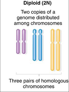
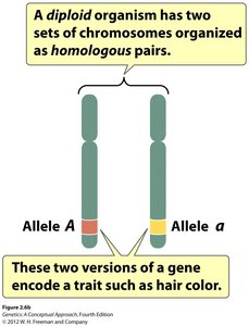
Chromosomes exist in pairs in diploid organisms, with one member from each parent. The complete set is called a karyotype.
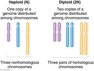
Diploid (2N): Two sets of chromosomes, one from each parent.
Haploid (N): One set of chromosomes, found in gametes.
Sex Chromosomes: Differ between males and females (e.g., XX in females, XY in males).
Autosomes: All other chromosomes not involved in sex determination.
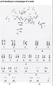
Example: Humans have 23 pairs of chromosomes (22 autosomes, 1 pair of sex chromosomes).
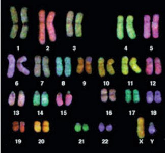
Karyotype and Chromosome Visualization
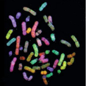
Karyotyping arranges chromosomes by size and shape to detect abnormalities and determine sex. Chromosomes can be visualized using staining techniques and fluorescent probes (FISH).
The Cell Cycle
Phases of the Cell Cycle
The cell cycle consists of four main phases: G1, S, G2, and M. Most of the cycle is spent in interphase, where chromosomes are not visible.
G1 Phase: Cell growth and preparation for DNA replication.
S Phase: DNA replication, creating sister chromatids.
G2 Phase: Preparation for mitosis.
M Phase: Mitosis and cytokinesis.
Mitosis
Purpose and Overview
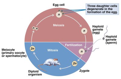
Mitosis is the process by which somatic cells divide, producing two identical daughter cells with the same chromosome number as the parent cell. It ensures genetic consistency across tissues.
Key Point: Chromosome number does not change during mitosis.
Key Point: Each daughter cell receives a maternal and paternal copy of each chromosome.
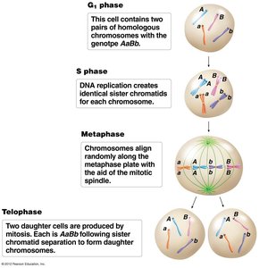
Stages of Mitosis
Mitosis is divided into several stages, each characterized by specific chromosomal and cellular events.
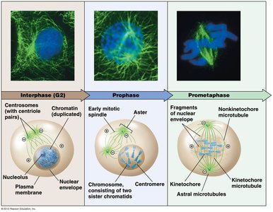
Interphase: Chromosomes are duplicated but not visible; nuclear envelope is intact.
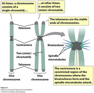
Prophase: Chromosomes condense and become visible as sister chromatids joined at the centromere.
Prometaphase: Nuclear envelope breaks down; spindle fibers attach to kinetochores.
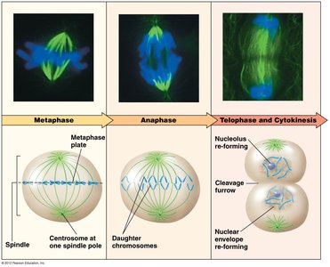
Metaphase: Chromosomes align at the metaphase plate.
Anaphase: Centromeres divide; sister chromatids are pulled to opposite poles.
Telophase: Nuclear envelope reforms; chromosomes decondense.
Cytokinesis: Cell divides into two.

Chromosome Structure During Mitosis
Sister Chromatids: Identical copies of a chromosome after DNA replication.
Homologous Chromosomes: Maternal and paternal copies, may carry different alleles.
Meiosis
Purpose and Overview
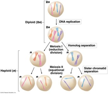
Meiosis is a specialized cell division that produces haploid gametes (sperm and egg) in sex organs. It involves one round of DNA replication followed by two cell divisions (meiosis I and II), resulting in four genetically distinct haploid cells.
Meiosis I: Reduction division; chromosome number is halved.
Meiosis II: Equational division; similar to mitosis.

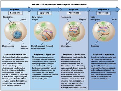
Stages of Meiosis I
Meiosis I is more complex than mitosis, especially during prophase I, which is subdivided into five stages.
Leptotene: Chromosomes become visible.
Zygotene: Synapsis or pairing of homologous chromosomes.
Pachytene: Chromosomes thicken; crossing over/recombination occurs.
Diplotene: Homologous chromosomes held together by chiasmata.
Diakinesis: Chromosomes fully condensed; nuclear envelope breaks down.
Meiosis II
Meiosis II separates sister chromatids, similar to mitosis, resulting in four haploid cells.
Mendel’s Laws and Chromosome Behavior
Law of Segregation
Each organism has two copies of each gene (alleles), which segregate randomly into gametes during meiosis. This explains why offspring inherit one allele from each parent.
Law of Independent Assortment
Alleles for different traits segregate independently if they are on different chromosomes. Chromosomes align randomly during metaphase I, leading to independent assortment.
Summary Table: Chromosome Types and Cell Division
Type | Chromosome Number | Cell Type | Division Process |
|---|---|---|---|
Diploid (2N) | Two sets | Somatic cells | Mitosis |
Haploid (N) | One set | Gametes | Meiosis |
Sex Chromosomes | XX or XY | Male/Female | Meiosis |
Autosomes | Pairs 1-22 | All cells | Mitosis/Meiosis |
Conclusion
The chromosome theory of inheritance provides the physical basis for Mendel’s laws, explaining how genes are transmitted through generations via the behavior of chromosomes during mitosis and meiosis. Understanding these processes is fundamental to genetics and heredity.
