Ch. 20 The Heart
Martini - Fundamentals of Anatomy & Physiology 11th Edition
Martini, Nath, Bartholomew11th EditionFundamentals of Anatomy & PhysiologyISBN: 9780136874089당신이 사용하는 게 아니라요?교과서 변경
20장, 문제 9

Identify the structures in the following diagram of a sectional view of the heart.
a. ___
b. ___
c. ___
d. ___
e. ___
f. ___
g. ___
h. ___
i. ___
j. ___
k. ___
l. ___
m. ___

검증된 단계별 안내
1
Step 1: Begin by carefully examining the sectional view of the heart provided in the diagram. Identify the major chambers first: the right atrium, right ventricle, left atrium, and left ventricle. These are the primary structural components of the heart.
Step 2: Next, locate the major valves that regulate blood flow between chambers and vessels. These include the tricuspid valve (between right atrium and right ventricle), the pulmonary valve (leading to the pulmonary artery), the mitral valve (between left atrium and left ventricle), and the aortic valve (leading to the aorta).
Step 3: Identify the major blood vessels connected to the heart. These typically include the superior and inferior vena cava (bringing deoxygenated blood to the right atrium), the pulmonary arteries (carrying blood from the right ventricle to the lungs), the pulmonary veins (bringing oxygenated blood to the left atrium), and the aorta (carrying oxygenated blood from the left ventricle to the body).
Step 4: Look for additional anatomical features such as the interventricular septum, which separates the left and right ventricles, and the interatrial septum, which separates the atria. These structures are important for understanding the heart's internal division.
Step 5: Assign each label (a through m) to the corresponding structure based on its position and characteristics in the diagram, using your knowledge of heart anatomy and the typical layout of these components in a sectional view.

비슷한 문제에 대한 검증된 영상 답변:

이 영상 해법은 위 문제에 도움이 된다고 튜터들이 추천한 것입니다.

주요 개념

질문에 올바르게 답하기 위해 반드시 이해해야 하는 핵심 개념들은 다음과 같습니다.

Anatomy of the Heart

Understanding the heart's anatomy is essential for identifying its structures in sectional views. This includes recognizing chambers (atria and ventricles), valves (tricuspid, mitral, pulmonary, aortic), and major vessels (aorta, pulmonary artery, vena cava). Familiarity with their relative positions helps in accurate labeling.
추천 영상:
6:13
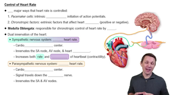
Control of Heart Rate

Sectional Views and Orientation

A sectional view slices through the heart to reveal internal structures. Knowing how to interpret these views requires understanding anatomical planes (sagittal, coronal, transverse) and orientation terms (anterior, posterior, left, right). This aids in correctly identifying which part of the heart is shown.
추천 영상:
02:33
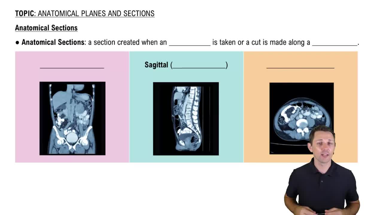
Anatomical Sections

Function and Flow of Blood Through the Heart

Comprehending the direction of blood flow through the heart chambers and valves clarifies the role of each structure. Blood flows from the body to the right atrium, right ventricle, lungs, left atrium, left ventricle, and then to the body. This functional knowledge supports accurate identification of heart parts.
추천 영상:
가이드 코스
6:11
Functions of Blood
관련 실천
교과서 질문

What role do the chordae tendineae and papillary muscles play in the normal function of the AV valves?

1322
views
교과서 질문

During diastole, a chamber of the heart:

(a) Relaxes and fills with blood

(b) Contracts and pushes blood into an adjacent chamber

(c) Experiences a sharp increase in pressure

(d) Reaches a pressure of approximately 120 mm Hg

976
views
교과서 질문

Identify the superficial structures in the following diagram of the heart.

a. ___

b. ___

c. ___

d. ___

e. ___

f. ___

g. ___

h. ___

783
views
교과서 질문

Cardiac output is equal to the:

(a) Difference between the end-diastolic volume and the end-systolic volume

(b) Product of heart rate and stroke volume

(c) Difference between the stroke volume at rest and the stroke volume during exercise

(d) Stroke volume less the end-systolic volume

(e) Product of heart rate and blood pressure

1164
views
교과서 질문

During the cardiac cycle, the amount of blood ejected from the left ventricle when the semilunar valve opens is the:

(a) Stroke volume (SV)

(b) End-diastolic volume (EDV)

(c) End-systolic volume (ESV)

(d) Cardiac output (CO)

1192
views
교과서 질문

The cardiac skeleton of the heart has which two of the following functions?

(a) It physically isolates the muscle fibers of the atria from those of the ventricles.

(b) It maintains the normal shape of the heart.

(c) It helps distribute the forces of cardiac contraction.

(d) It allows more rapid contraction of the ventricles.

(e) It strengthens and helps prevent overexpansion of the heart.

1010
views