Ch. 20 The Heart
Martini - Fundamentals of Anatomy & Physiology 11th Edition
Martini, Nath, Bartholomew11th EditionFundamentals of Anatomy & PhysiologyISBN: 9780136874089당신이 사용하는 게 아니라요?교과서 변경
20장, 문제 8

Identify the superficial structures in the following diagram of the heart.
a. ___
b. ___
c. ___
d. ___
e. ___
f. ___
g. ___
h. ___

검증된 단계별 안내
1
Step 1: Understand that superficial structures of the heart refer to the external anatomical features visible on the heart's surface, such as major vessels, grooves, and chambers.
Step 2: Identify the major external vessels typically labeled in heart diagrams, including the aorta, pulmonary trunk, superior vena cava, and inferior vena cava.
Step 3: Locate the coronary sulcus (atrioventricular groove) and the anterior interventricular sulcus, which are grooves on the heart's surface marking boundaries between chambers.
Step 4: Recognize the visible chambers on the heart's surface, such as the right atrium, right ventricle, left atrium, and left ventricle, based on their position relative to the grooves and vessels.
Step 5: Match each label (a through h) to these superficial structures by comparing their position on the diagram to the known anatomical landmarks and vessels described.

비슷한 문제에 대한 검증된 영상 답변:

이 영상 해법은 위 문제에 도움이 된다고 튜터들이 추천한 것입니다.

주요 개념

질문에 올바르게 답하기 위해 반드시 이해해야 하는 핵심 개념들은 다음과 같습니다.

Anatomy of the Heart's Superficial Structures

Superficial structures of the heart refer to the external features visible on its surface, such as the atria, ventricles, coronary sulcus, and major blood vessels. Recognizing these helps in identifying parts labeled in diagrams and understanding heart function and blood flow.
추천 영상:
6:13
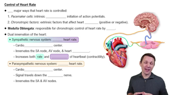
Control of Heart Rate

Coronary Sulcus and Interventricular Sulci

The coronary sulcus is a groove separating the atria from the ventricles, while the anterior and posterior interventricular sulci separate the left and right ventricles. These sulci contain important blood vessels and are key landmarks in heart surface anatomy.
추천 영상:
5:10
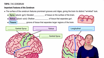
Important Features of the Cerebrum

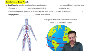
Major Blood Vessels Associated with the Heart

The aorta, pulmonary trunk, superior and inferior vena cava, and pulmonary veins are major vessels connected to the heart's surface. Identifying these vessels is essential for understanding the heart’s role in systemic and pulmonary circulation.
추천 영상:
관련 실천
교과서 질문

The heart is surrounded by the:

(a) Pleural cavity

(b) Peritoneal cavity

(c) Abdominopelvic cavity

(d) Mediastinum

(e) Abdominal cavity

550
views
교과서 질문

Identify the structures in the following diagram of a sectional view of the heart.

a. ___

b. ___

c. ___

d. ___

e. ___

f. ___

g. ___

h. ___

i. ___

j. ___

k. ___

l. ___

m. ___

885
views
교과서 질문

During diastole, a chamber of the heart:

(a) Relaxes and fills with blood

(b) Contracts and pushes blood into an adjacent chamber

(c) Experiences a sharp increase in pressure

(d) Reaches a pressure of approximately 120 mm Hg

976
views
교과서 질문

Cardiac output is equal to the:

(a) Difference between the end-diastolic volume and the end-systolic volume

(b) Product of heart rate and stroke volume

(c) Difference between the stroke volume at rest and the stroke volume during exercise

(d) Stroke volume less the end-systolic volume

(e) Product of heart rate and blood pressure

1164
views
교과서 질문

During the cardiac cycle, the amount of blood ejected from the left ventricle when the semilunar valve opens is the:

(a) Stroke volume (SV)

(b) End-diastolic volume (EDV)

(c) End-systolic volume (ESV)

(d) Cardiac output (CO)

1192
views
교과서 질문

The cardiac skeleton of the heart has which two of the following functions?

(a) It physically isolates the muscle fibers of the atria from those of the ventricles.

(b) It maintains the normal shape of the heart.

(c) It helps distribute the forces of cardiac contraction.

(d) It allows more rapid contraction of the ventricles.

(e) It strengthens and helps prevent overexpansion of the heart.

1010
views