Ch. 4 The Tissue Level of Organization
Martini - Fundamentals of Anatomy & Physiology 11th Edition
Martini, Nath, Bartholomew11th EditionFundamentals of Anatomy & PhysiologyISBN: 9780136874089당신이 사용하는 게 아니라요?교과서 변경
4장, 문제 31

During a lab practical, a student examines a tissue that is composed of densely packed protein fibers that run parallel to each other and form a cord. There are no striations, but small nuclei are visible. The student identifies the tissue as skeletal muscle. Why is the student's choice wrong, and what tissue is he probably observing?

검증된 단계별 안내
1
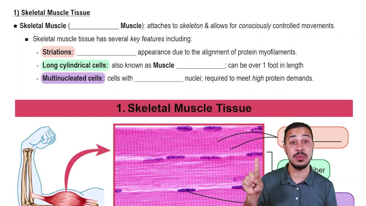
Step 1: Recall the characteristics of skeletal muscle tissue. Skeletal muscle fibers are long, cylindrical, multinucleated cells with obvious striations (alternating light and dark bands) due to the organized arrangement of actin and myosin filaments.
Step 2: Note that the problem states the tissue has densely packed protein fibers running parallel, no striations, and small visible nuclei. The absence of striations is a key clue that this is not skeletal muscle, since skeletal muscle always shows striations under the microscope.
Step 3: Consider other types of muscle tissue. Smooth muscle tissue also has densely packed fibers running parallel, but unlike skeletal muscle, smooth muscle fibers are spindle-shaped with a single, centrally located nucleus and no striations.
Step 4: Recognize that the description fits smooth muscle tissue, which is found in walls of hollow organs and blood vessels, and appears as densely packed, non-striated fibers with small nuclei.
Step 5: Conclude that the student's identification is incorrect because the tissue lacks striations, and the tissue observed is most likely smooth muscle, not skeletal muscle.

비슷한 문제에 대한 검증된 영상 답변:

이 영상 해법은 위 문제에 도움이 된다고 튜터들이 추천한 것입니다.
영상 길이:
5m
도움이 되었나요?

주요 개념

질문에 올바르게 답하기 위해 반드시 이해해야 하는 핵심 개념들은 다음과 같습니다.

Skeletal Muscle Tissue Characteristics

Skeletal muscle tissue is composed of long, cylindrical fibers that are multinucleated and exhibit visible striations due to the organized arrangement of actin and myosin filaments. These striations are a key identifying feature, and the nuclei are typically located at the periphery of the fibers.
추천 영상:
가이드 코스
06:56
Skeletal Muscle Tissue

Dense Regular Connective Tissue Structure

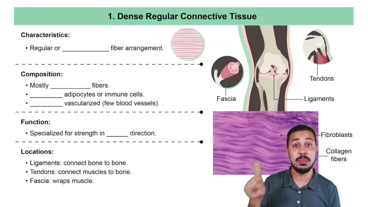
Dense regular connective tissue consists of tightly packed collagen fibers arranged in parallel bundles, providing great tensile strength. It lacks striations and contains small, flattened nuclei of fibroblasts scattered between the fibers, which can be mistaken for muscle nuclei under the microscope.
추천 영상:
가이드 코스
06:29
Dense Regular Connective Tissue

Histological Differences Between Muscle and Connective Tissue

Muscle tissues show striations and organized contractile proteins, while connective tissues like tendons have collagen fibers without striations. Recognizing the presence or absence of striations and the shape and position of nuclei helps differentiate these tissues during microscopic examination.
추천 영상:
가이드 코스
08:25
Areolar Connective Tissue