Pearson+ LogoPearson+ Logo
Ch. 10 - Eukaryotic Chromosome Abnormalities and Molecular Organization
Sanders - Genetic Analysis: An Integrated Approach 3rd Edition
Sanders3rd EditionGenetic Analysis: An Integrated ApproachISBN: 9780135564172Not the one you use?Change textbook
Chapter 10, Problem 16c

The accompanying chromosome diagram represents a eukaryotic chromosome prepared with Giemsa stain. Indicate the heterochromatic and euchromatic regions of the chromosome, and label the chromosome's centromeric and telomeric regions.

Why are expressed genes not found in the telomeric region of chromosomes?

Verified step by step guidance
1
Examine the chromosome diagram and identify the darker-stained regions. These darker regions represent heterochromatin, which is tightly packed DNA and generally transcriptionally inactive.
Identify the lighter-stained regions on the chromosome. These lighter regions represent euchromatin, which is loosely packed DNA and typically transcriptionally active.
Locate the centromeric region of the chromosome. This is the constricted area that divides the chromosome into two arms and is essential for proper chromosome segregation during cell division.
Identify the telomeric regions at the ends of the chromosome. These are specialized structures that protect the ends of the chromosome from degradation and prevent them from fusing with other chromosomes.
Explain why expressed genes are not found in the telomeric region: Telomeric regions consist of repetitive DNA sequences and are highly condensed, forming heterochromatin. This structure is not conducive to gene expression because the transcriptional machinery cannot easily access the DNA in these regions.

Verified video answer for a similar problem:

This video solution was recommended by our tutors as helpful for the problem above.
Video duration:
2m
Was this helpful?

Key Concepts

Here are the essential concepts you must grasp in order to answer the question correctly.

Euchromatin and Heterochromatin

Euchromatin is a less condensed form of chromatin that is actively involved in transcription, allowing genes to be expressed. In contrast, heterochromatin is tightly packed and generally transcriptionally inactive, serving structural roles in the chromosome. Understanding the distinction between these two types of chromatin is essential for identifying regions of gene expression.
Recommended video:

Telomeres

Telomeres are repetitive nucleotide sequences located at the ends of chromosomes that protect them from degradation and prevent fusion with neighboring chromosomes. They play a crucial role in maintaining chromosome stability but are not involved in gene expression, which is why expressed genes are typically absent in these regions.
Recommended video:
Guided course
08:38
Telomeres and Telomerase

Centromeres

The centromere is the region of a chromosome where the two sister chromatids are joined and is essential for proper chromosome segregation during cell division. It is characterized by specific DNA sequences and proteins that facilitate the attachment of spindle fibers. While centromeres are critical for chromosome function, they also do not contain actively expressed genes.
Recommended video:
Guided course
04:38
Rearrangement Overview
Related Practice
Textbook Question

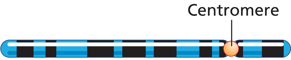
A normal chromosome and its homolog carrying a paracentric inversion are shown here. The dot (·) represents the centromere.

Normal    ABC • DEFGHIJK

Inversion  abc • djihgfe

Assume a crossover takes place in the region between A and B. Identify the gametes that are formed by this crossover event, and indicate which, if any, gametes are viable.

462
views
Textbook Question

The accompanying chromosome diagram represents a eukaryotic chromosome prepared with Giemsa stain. Indicate the heterochromatic and euchromatic regions of the chromosome, and label the chromosome's centromeric and telomeric regions.

What term best describes the shape of this chromosome?

527
views
Textbook Question

The accompanying chromosome diagram represents a eukaryotic chromosome prepared with Giemsa stain. Indicate the heterochromatic and euchromatic regions of the chromosome, and label the chromosome's centromeric and telomeric regions.

Do you expect the centromeric region to contain heterochromatin? Why or why not?

477
views
Textbook Question

The accompanying chromosome diagram represents a eukaryotic chromosome prepared with Giemsa stain. Indicate the heterochromatic and euchromatic regions of the chromosome, and label the chromosome's centromeric and telomeric regions.

Are you more likely to find the DNA sequence encoding the digestive enzyme amylase in a heterochromatic, euchromatic, centromeric, or telomeric region? Explain your reasoning.

561
views
Textbook Question

Histone protein H4 isolated from pea plants and cow thymus glands contains 102 amino acids in both cases. A total of 100 of the amino acids are identical between the two species. Give an evolutionary explanation for this strong amino acid sequence identity based on what you know about the functions of histones and nucleosomes.

475
views
Textbook Question

A survey of organisms living deep in the ocean reveals two new species whose DNA is isolated for analysis. DNA samples from both species are treated to remove nonhistone proteins. Each DNA sample is then treated with DNase I that cuts DNA not protected by histone proteins but is unable to cut DNA bound by histone proteins. Following DNase I treatment, DNA samples are subjected to gel electrophoresis, and the gels are stained to visualize all DNA bands in the gel. The staining patterns of DNA bands from each species are shown in the figure. The number of base pairs in small DNA fragments is shown at the left of the gel. Interpret the gel results in terms of chromatin organization and the spacing of nucleosomes in the chromatin of each species.

450
views