Pearson+ LogoPearson+ Logo
Ch. 15 - Recombinant DNA Technology and Its Applications
Sanders - Genetic Analysis: An Integrated Approach 3rd Edition
Sanders3rd EditionGenetic Analysis: An Integrated ApproachISBN: 9780135564172Not the one you use?Change textbook
Chapter 15, Problem 18

To further analyze the CRABS CLAW gene, you create a map of the genomic clone. The 11-kb EcoRI fragment is ligated into the EcoRI site of the MCS of the vector shown in Problem 18. You digest the double-stranded form of the genome with several restriction enzymes and obtain the following results. Draw, as far as possible, a map of the genomic clone of CRABS CLAW.
Table displaying restriction enzyme digestion results for the CRABS CLAW gene, including fragment sizes in kilobases.
What restriction digest would help resolve any ambiguity in the map?

Verified step by step guidance
1
Step 1: Begin by analyzing the data provided for each restriction enzyme digest. Note the fragment sizes generated by EcoRI, XbaI, XhoI, SalI, and HindIII individually, as well as the combinations of EcoRI with each enzyme. This will help identify overlapping fragments and potential cut sites.
Step 2: Construct a preliminary map of the genomic clone based on the fragment sizes. Start with the EcoRI digest, which produces fragments of 11.0 kb and 3.0 kb. Place these fragments on the map, ensuring their total length matches the genomic clone size.
Step 3: Incorporate the results of the double digests (e.g., EcoRI + XbaI, EcoRI + XhoI, etc.) into the map. For example, the EcoRI + XbaI digest produces fragments of 4.5 kb, 6.5 kb, and 3.0 kb. Use this information to determine the relative positions of XbaI cut sites within the EcoRI fragments.
Step 4: Repeat the process for the other double digests (EcoRI + XhoI, EcoRI + SalI, EcoRI + HindIII). Compare the fragment sizes generated by each combination to refine the map and resolve ambiguities in the placement of restriction sites.
Step 5: To resolve any remaining ambiguities, consider performing additional restriction digests with enzymes not yet used or combinations of enzymes that target specific regions of the map. For example, a digest with XbaI + XhoI or SalI + HindIII could provide further clarity on overlapping fragments or closely spaced cut sites.

Verified video answer for a similar problem:

This video solution was recommended by our tutors as helpful for the problem above.
Video duration:
2m
Was this helpful?

Key Concepts

Here are the essential concepts you must grasp in order to answer the question correctly.

Restriction Enzymes

Restriction enzymes, or restriction endonucleases, are proteins that cut DNA at specific sequences, allowing scientists to manipulate and analyze genetic material. Each enzyme recognizes a unique sequence of nucleotides, which is crucial for creating DNA fragments of desired lengths for cloning or mapping. Understanding how these enzymes work is essential for interpreting the results of a restriction digest and constructing a genomic map.
Recommended video:
Guided course
07:11
Mapping with Markers

Genomic Mapping

Genomic mapping involves determining the locations of genes and other important sequences on a chromosome. This process often utilizes restriction enzyme digests to create a physical map of DNA fragments, which can be analyzed to understand gene organization and function. A well-constructed map helps resolve ambiguities in gene locations and relationships, making it a fundamental aspect of genetic analysis.
Recommended video:
Guided course
03:04
Modern Mapping

Fragment Size Analysis

Fragment size analysis is a technique used to determine the lengths of DNA fragments generated by restriction enzyme digestion. By comparing the sizes of these fragments, researchers can infer the positions of restriction sites and the overall structure of the genomic clone. This analysis is critical for resolving ambiguities in genomic maps, as different combinations of restriction enzymes can yield distinct patterns that clarify the arrangement of genetic elements.
Recommended video:
Guided course
02:48
Chi Square Analysis
Related Practice
Textbook Question

The bacteriophage lambda genome can exist in either a linear form or a circular form.

Diagram the resulting fragments as they would appear on an agarose gel after electrophoresis.

453
views
Textbook Question

The restriction enzymes XhoI and SalI cut their specific sequences as shown below:

Can the sticky ends created by XhoI and SalI sites be ligated? If yes, can the resulting sequences be cleaved by either XhoI or SalI?

1561
views
Textbook Question

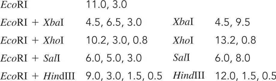
The bacteriophage ϕX174 has a single-stranded DNA genome of 5386 bases. During DNA replication, double-stranded forms of the genome are generated. In an effort to create a restriction map of ϕX174, you digest the z-stranded form of the genome with several restriction enzymes and obtain the following results. Draw a map of the ϕX174 genome.

645
views
Textbook Question

You have isolated a genomic clone with an EcoRI fragment of 11 kb that encompasses the CRABS CLAW gene. You digest the genomic clone with HindIII and note that the 11-kb EcoRI fragment is split into three fragments of 9 kb, 1.5 kb, and 0.5 kb.

Does this tell you anything about where the CRABS CLAW gene is located within the 11-kb genomic clone?

612
views
Textbook Question

You have isolated a genomic clone with an EcoRI fragment of 11 kb that encompasses the CRABS CLAW gene (see Problem 18). You digest the genomic clone with HindIII and note that the 11-kb EcoRI fragment is split into three fragments of 9 kb, 1.5 kb, and 0.5 kb.

Restriction enzyme sites within a cDNA clone are often also found in the genomic sequence. Can you think of a reason why occasionally this is not the case? What about the converse: Are restriction enzyme sites in a genomic clone always in a cDNA clone of the same gene?

553
views
Textbook Question

You have identified a 0.80-kb cDNA clone that contains the entire coding sequence of the Arabidopsis gene CRABS CLAW. In the construction of the cDNA library, linkers with EcoRI sites were added to each end of the cDNA, and the cDNA was inserted into the EcoRI site of the MCS of the vector shown in the accompanying figure. You perform digests on the CRABS CLAW cDNA clone with restriction enzymes and obtain the following results. Can you determine the orientation of the cDNA clone with respect to the restriction enzyme sites in the vector? The restriction enzyme sites listed in the dark blue region are found only in the MCS of the vector.

875
views