Pearson+ LogoPearson+ Logo
Ch. 8 - Molecular Biology of Transcription and RNA Processing
Sanders - Genetic Analysis: An Integrated Approach 3rd Edition
Sanders3rd EditionGenetic Analysis: An Integrated ApproachISBN: 9780135564172Not the one you use?Change textbook
Chapter 8, Problem 18c

A 3.5-kb segment of DNA containing the complete sequence of a mouse gene is available. The DNA segment contains the promoter sequence and extends beyond the polyadenylation site of the gene. The DNA is studied by band shift assay, and the following gel bands are observed.
Gel electrophoresis image showing five lanes with distinct bands representing a 3.5-kb DNA fragment.
Match these conditions to a specific lane of the gel.
3.5-kb fragment alone

Verified step by step guidance
1
Step 1: Understand the context of the problem. A band shift assay is used to study DNA-protein interactions. In this case, the 3.5-kb DNA fragment contains the complete sequence of a mouse gene, including the promoter and polyadenylation site. The gel bands observed represent different conditions of the DNA fragment being studied.
Step 2: Focus on the condition described: '3.5-kb fragment alone.' This means the DNA fragment is not interacting with any proteins or other molecules. It is simply the isolated DNA segment being analyzed.
Step 3: Recall that in a band shift assay, the DNA fragment alone will migrate through the gel based on its size and charge. Since no proteins or other molecules are bound to the DNA, its migration will be unaltered, and it will appear as a single band corresponding to its size.
Step 4: Match this condition to the specific lane of the gel. The lane representing the '3.5-kb fragment alone' will show a single band at the position corresponding to the size of the DNA fragment (3.5 kb). This is typically the control lane in the assay.
Step 5: To confirm, compare the migration pattern of the DNA fragment alone to other lanes where DNA-protein interactions may be present. The '3.5-kb fragment alone' lane should have the simplest pattern, with no shifts or additional bands.

Verified video answer for a similar problem:

This video solution was recommended by our tutors as helpful for the problem above.
Video duration:
1m
Was this helpful?

Key Concepts

Here are the essential concepts you must grasp in order to answer the question correctly.

Promoter Sequence

The promoter sequence is a region of DNA located upstream of a gene that initiates transcription. It contains specific binding sites for RNA polymerase and transcription factors, which are essential for the regulation of gene expression. Understanding the promoter's role is crucial for interpreting how the gene is activated or silenced in response to various signals.
Recommended video:
Guided course
08:41
Sequencing Difficulties

Polyadenylation Site

The polyadenylation site is a sequence in the mRNA that signals the addition of a poly(A) tail, which is important for mRNA stability, export from the nucleus, and translation efficiency. In the context of the DNA segment, knowing where the polyadenylation site is located helps in understanding the full length of the mRNA produced from the gene and its functional implications.
Recommended video:
Guided course
08:39
mRNA Processing

Band Shift Assay

A band shift assay, also known as an electrophoretic mobility shift assay (EMSA), is a technique used to study protein-DNA interactions. In this assay, DNA fragments bound to proteins migrate more slowly through a gel than unbound DNA, resulting in distinct bands. This method is essential for analyzing how transcription factors interact with the promoter region of the gene in question.
Recommended video:
Guided course
03:44
Plaques and Experiments
Related Practice
Textbook Question

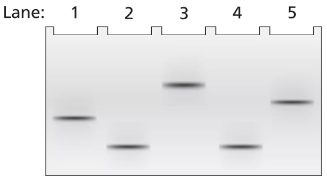
A 2-kb fragment of E. coli DNA contains the complete sequence of a gene for which transcription is terminated by the rho protein. The fragment contains the complete promoter sequence as well as the terminator region of the gene. The cloned fragment is examined by band shift assay. Each lane of a single electrophoresis gel contains the 2-kb cloned fragment under the following conditions:

Lane 1: 2-kb fragment alone

Lane 2: 2-kb fragment plus the core enzyme

Lane 3: 2-kb fragment plus the RNA polymerase holoenzyme

Lane 4: 2-kb fragment plus rho protein

Explain the relative positions of bands in lanes 1 and 4.

504
views
Textbook Question

A 3.5-kb segment of DNA containing the complete sequence of a mouse gene is available. The DNA segment contains the promoter sequence and extends beyond the polyadenylation site of the gene. The DNA is studied by band shift assay, and the following gel bands are observed.

Match these conditions to a specific lane of the gel.

3.5-kb fragment plus TFIIB and TFIID

372
views
Textbook Question

A 3.5-kb segment of DNA containing the complete sequence of a mouse gene is available. The DNA segment contains the promoter sequence and extends beyond the polyadenylation site of the gene. The DNA is studied by band shift assay, and the following gel bands are observed.

Match these conditions to a specific lane of the gel.

3.5-kb fragment plus TFIIB, TFIID, TFIIF, and RNA polymerase II

374
views
Textbook Question

A 3.5-kb segment of DNA containing the complete sequence of a mouse gene is available. The DNA segment contains the promoter sequence and extends beyond the polyadenylation site of the gene. The DNA is studied by band shift assay, and the following gel bands are observed.

Match these conditions to a specific lane of the gel.

3.5-kb fragment plus RNA polymerase II

354
views
Textbook Question

A 3.5-kb segment of DNA containing the complete sequence of a mouse gene is available. The DNA segment contains the promoter sequence and extends beyond the polyadenylation site of the gene. The DNA is studied by band shift assay, and the following gel bands are observed.

Match these conditions to a specific lane of the gel.

3.5-kb fragment plus TFIIB

473
views
Textbook Question

A 1.0-kb DNA fragment from the end of the mouse gene described in the previous problem is examined by DNA footprint protection analysis. Two samples are end-labeled with ³²P and one of the two is mixed with TFIIB, TFIID, and RNA polymerase II. The DNA exposed to these proteins is run in the right-hand lane of the gel shown below and the control DNA is run in the left-hand. Both DNA samples are treated with DNase I before running the samples on the electrophoresis gel.

What length of DNA is bound by the transcriptional proteins? Explain how the gel results support this interpretation.

559
views