Pearson Interactive Labs
Microbiology
Reimagining online labs
Microbiology
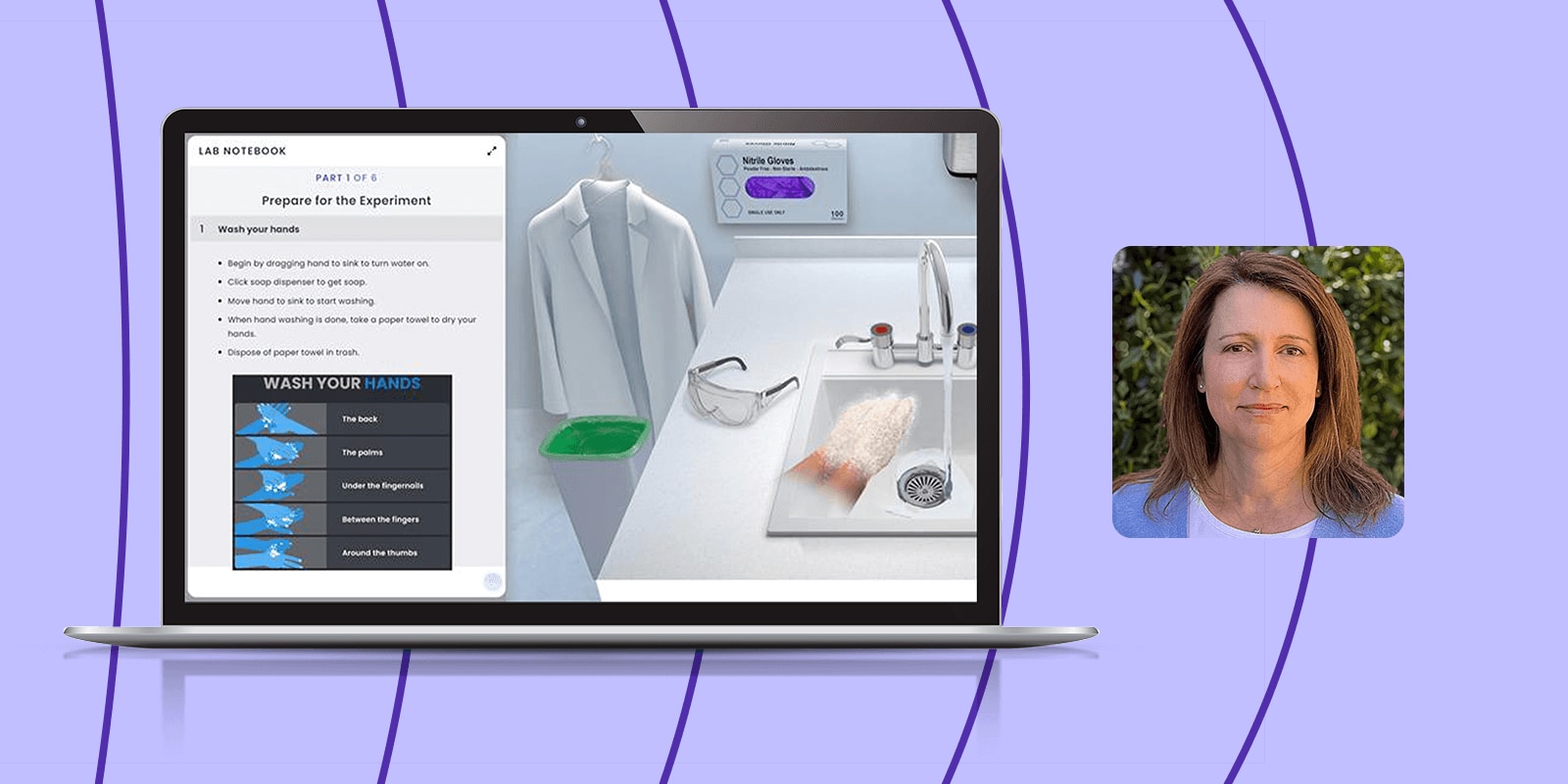
Pearson® Interactive Labs for Microbiology is an easy-to-use suite of online microbiology lab simulations. Real-world clinical scenarios create an immersive experience where students learn by doing. Students receive guided feedback as they master lab techniques. All labs include customizable post-lab assessment. You can access the following labs with Pearson Microbiology titles in Mastering® Microbiology at no additional cost. Pearson Microbiology titles also include access to biology labs.
“The authentic lab design simulates hands-on procedures realistically, including correct handling of equipment and error recognition, creating meaningful engagement.”
- 2025 CODiE Award Judge

Pearson Interactive Labs is a 2025 CODiE Award winner in the Best Science Instructional Solution category.
The CODiE Awards represent the very best in technology. Recognition as a winner demonstrates excellence, impact, and leadership in driving the future of technology.

A Comprehensive Lab Experience: The Journey to Creating Pearson Interactive Labs
Author Dr. Katherine Rawls shares her process behind creating complete virtual Microbiology labs that integrate techniques, concepts, and analysis and promote active learning alongside career relevance.
Gram Stain
Students conduct a Gram stain analysis of a bacterial culture to support a diagnosis and antibiotic treatment. When the slide is visualized under the microscope, students use the control smears to diagnose how decolorization went. They sort out challenges with Gram staining, including mistakes with decolorization, use of an aging culture, and interpretation of control smears.
Lab content includes:
Methods/Techniques
- Decolorization
- Heat-fixing a smear
- Stain and counterstain application
- Lab safety and disinfection protocols
Staining
- Gram stain
Concepts
- Cell anatomy
- Gram-positive and Gram-negative bacteria
Analysis/Interpretation
- Results of Gram-stained specimens
- Results of stains
- Errors and misconceptions
- Clinical applications
NEW! Biochemical Tests: Gram Positive
In this virtual laboratory exercise, students will use biochemical tests to characterize an unknown Gram-positive bacterium, including the catalase test, phenyl ethyl alcohol agar, mannitol salt agar, blood agar, DNase agar and bile esculin agar.
Lab content includes:
Tests/Media
- Bile Esculin agar
- Blood agar (hemolytic patterns)
- Catalase test
- DNase agar
- Mannitol salt agar (mannitol fermentation and halotolerance)
- Phenylethyl Alcohol agar
Methods/Techniques
- Solid culture to nutrient agar plate
- Lab safety and disinfection protocols
Concepts
- Infection identification through tests
- Selective and differential media
- Unknown specimen identification
- Use of controls
Analysis/Interpretation
- Results of biochemical tests
- Errors and misconceptions
- Clinical applications
Aseptic Technique
Students use aseptic technique to transfer a bacterial culture to various media without contamination. They work progressively through the steps of aseptic technique using three culture media. While transferring from the first media, student errors are identified and corrected. During the second transfer, students have to rely on what they learned previously to make decisions, but their errors are caught and corrected. In the final transfer, they must recall the steps of aseptic technique and apply them.
Lab content includes:
Methods/Techniques
- Liquid culture to nutrient agar plate
- Liquid culture to nutrient agar slant
- Liquid culture to nutrient broth
- Lab safety and disinfection protocols
Concepts
- Aseptic transfer
- Growth media
Analysis/Interpretation
- Results of successful/unsuccessful aseptic transfer
- Errors and misconceptions
- Clinical applications
Streak for Isolation
In this virtual lab, students will perform a streak for isolation to isolate different types of bacteria that are present in a sample from an infected wound. The isolation streaks are performed to obtain single, isolated colonies that will be further differentiated by the colony characteristics. The isolation streak will separate out the infecting bacteria as pure colonies, which can be sampled and identified, helping inform treatment.
Lab content includes:
Methods/Techniques
- T-streak method
- Quadrant-streak method
- Lab safety and disinfection protocols
Concepts
- Bacterial colonies
- Isolation streak
Analysis/Interpretation
- Results of isolation streaks
- Errors and misconceptions
- Clinical applications
Acid-Fast Stain
In this virtual lab, students will perform acid-fast stains on control organisms and then analyze them along with an acid-fast stain from a sick patient’s sputum sample to determine the presence of acid-fast bacteria. Students will gain an understanding of the benefits of the acid-fast stain as a reliable, rapid detection technique for acid-fast bacteria, like the one that causes tuberculosis.
Lab content includes:
Methods/Techniques
- Decolorization
- Stain and counterstain application
- Lab safety and disinfection protocols
Staining
- Acid-fast stain (Ziehl-Neelsen) method
Concepts
- Cell anatomy
- Infection identification through tests
Analysis/Interpretation
- Results of stains
- Errors and misconceptions
- Clinical applications
Antimicrobial Susceptibility Techniques
Students will become well-versed in the Kirby-Bauer antimicrobial sensitivity procedure in this virtual lab. After a procedural overview on how to perform and interpret results, students will perform the procedure on two pure liquid bacterial cultures and evaluate drug sensitivity based on measured zones of inhibition and their correlation with values in a standard table. The lesson ends by examining the clinical relevance of the learned technique as well as common misconceptions and technique errors.
Lab content includes:
Tests/Media
- Kirby-Bauer test
- Mueller-Hinton agar
Methods/Techniques
- Measuring zones of inhibition
- Plating bacteria using a cotton swab
- Lab safety and disinfection protocols
Concepts
- Antimicrobial drug zone diameters
Analysis/Interpretation
- Antimicrobial drug resistance
- Errors and misconceptions
- Clinical applications
NEW! Chemical Control of Microbial Growth
In this virtual lab, students will explore the effects of exposure time and gram type on the effectiveness of chemical control. Students will then analyze data to answer questions regarding the best chemicals to use in different situations and the success of various chemical treatments, including the treatment of Streptococcus mutans with mouthwash.
Lab content includes:
Tests/Media
- Dey-Engley agar
Methods/Techniques
- Plating bacteria using a cotton swab
- Using a serological pipette
- Lab safety and disinfection protocols
Concepts
- Colony counting
- Control of microbial growth
Analysis/Interpretation
- Errors and misconceptions
- Clinical applications
NEW! Biochemical Tests: Gram Negative
In this virtual laboratory exercise, students will use biochemical tests to characterize an unknown Gram-negative bacterium, including MacConkey agar, SIM agar, MRVP broth, urea broth, citrate agar, nitrate broth, and phenol red carbohydrate broth.
Lab content includes:
Tests/Media
- Citrate agar
- MacConkey agar
- Methyl Red – Voges Proskauer (MRVP) broth
- Nitrate broth
- Phenol red carbohydrate broth
- Sulfur indole motility (SIM) agar
- Urea broth
Methods/Techniques
- Adding test reagents
- Agar slant to broth media
- Solid culture to nutrient agar plate
- Lab safety and disinfection protocols
Concepts
- Infection identification through tests
- Selective and differential media
- Unknown specimen identification
- Use of controls
Analysis/Interpretation
- Results of biochemical tests
- Errors and misconceptions
- Clinical applications
Smear Prep
In this virtual lab, students prepare smears and perform simple stains on both solid and liquid bacterial cultures. They view stained specimens under the microscope to determine microbial presence and cellular morphology. Next, relying on what they have learned in the lab, students explore simple staining for presumptive pathogen identification. Finally, students will examine common errors like incorrect smear thickness and improper heat-fixing technique.
Lab content includes:
Methods/Techniques
- Heat-fixing a smear
- Smear prepping from broth and solid cultures
- Lab safety and disinfection protocols
Staining
- Simple stain from broth and solid cultures
Concepts
- Acidic and basic dyes
- Cellular morphology
- Staining/smear preparation
Analysis/Interpretation
- Errors and misconceptions
- Clinical applications
Microscopy (for Microbiology)
In this virtual lab experiment, students will learn the proper care of a microscope, its principal components, the common problems that can occur while in use and how to examine microbial samples to determine results. Students begin by visualizing the causative agent of a patient’s illness by bringing the culprit organism into focus and ultimately identifying the organism implicated by a rapid test.
Lab content includes:
Methods/Techniques
- Using a compound light microscope
- Lab safety and disinfection protocols
Concepts
- Calculation of total magnification
- Oil immersion
Analysis/Interpretation
- Errors and misconceptions
- Clinical applications
Endospore Stain
In this virtual lab, endospore stains will be performed on various bacterial specimens to identify the presence or absence of endospores. During this exercise, students will learn the process of endospore staining and apply this technique to control clinical samples. Next, they will conduct an endospore staining procedure to look for the presence of C. difficile under a microscope. This information will inform treatment and protect the other residents in the medical facility.
Lab content includes:
Methods/Techniques
- Decolorization
- Stain and counterstain application
- Lab safety and disinfection protocols
Staining
- Endospore stain
Concepts
- Endospores and vegetative cells
Analysis/Interpretation
- Results of stains
- Errors and misconceptions
- Clinical applications
NEW! Serial Dilution and Enumeration
In this virtual lab, students will explore the importance of using serial dilutions and viable plate counts to enumerate bacterial concentrations in real life samples. Students will also demonstrate the process of using the serial dilution and viable plate count to enumerate bacterial concentrations in clinical samples, thereby helping diagnose a simulated patient.
Lab content includes:
Methods/Techniques
- Spread plate method
- Serial dilution
- Using a micropipette
- Using an L-shaped spreader
- Lab safety and disinfection protocols
Concepts
- Bacteria concentration calculation
- Colony counting
- Viable plate count
Analysis/Interpretation
- Errors and misconceptions
- Clinical applications
NEW! Physical Control of Microbial Growth
In this virtual lab, students will explore the differences between various physical methods for their effectiveness at controlling the growth of microorganisms. Students will work as a member of a patient safety team to actively examine the efficacy of the use of dry heat, autoclave, and UV light as physical methods of controlling the growth of bacteria in a simulated clinical environment.
Lab content includes:
Methods/Techniques
- Plating bacteria using a cotton swab
- Using a dry-heat oven and autoclave
- Lab safety and disinfection protocols
Concepts
- Colony counting
- Control of microbial growth
Analysis/Interpretation
- Errors and misconceptions
- Clinical applications
NEW! Identifying the Unknown Mystery Microbe 1
In this virtual lab, students will identify an unknown mystery microbe to reinforce critical thinking and diagnostic skills in microbial identification. Using knowledge and techniques from previous labs, they will perform a Gram stain along with a series of biochemical tests to determine the genus and species of the microorganism responsible for a specific illness.
Lab content includes:
Methods/Techniques
- Adding test reagents
- Agar slant to broth media
- Solid culture to nutrient agar plate
- Lab safety and disinfection protocols
Concepts
- Use of controls
- Dichotomous key
- Selective and differential media
- Unknown specimen identification
- Infection identification through tests
Analysis/Interpretation
- Results of biochemical tests
- Results of Gram-stained specimens
- Errors and misconceptions
- Clinical applications
Tests/Media
- Citrate agar
- MacConkey agar
- Methyl Red – Voges Proskauer (MRVP) broth
- Ornithine decarboxylase broth
- Oxidase test
- Sulfur indole motility (SIM) agar
- Urea broth
Staining
- Gram stain
NEW! Identifying the Unknown Mystery Microbe 2
Lab content includes:
In the second virtual lab of our “Identifying the Unknown Microbe” series, students will identify a different unknown mystery microbe to reinforce critical thinking and diagnostic skills in microbial identification. Using knowledge and techniques from previous labs, they will perform a Gram stain along with a series of biochemical tests to determine the genus and species of the microorganism responsible for a specific illness.
Methods/Techniques
- Solid culture to nutrient agar plate
- Lab safety and disinfection protocols
Concepts
- Dichotomous key
- Infection identification through tests
- Selective and differential media
- Unknown specimen identification
- Use of controls
Analysis/Interpretation
- Results of biochemical tests
- Results of Gram-stained specimens
- Errors and misconceptions
- Clinical applications
Tests/Media
- Catalase test
- Coagulase test
- DNase agar
- Mannitol salt agar (mannitol fermentation and halotolerance)
Staining
- Gram stain

Transforming the Lab Experience with Pearson Interactive Labs for Microbiology
Join Dr. Donna Cain, professor of microbiology and author on Pearson Interactive Labs, as she shares ways to incorporate virtual wet lab experiences into your course. Discover strategies to enhance engagement and learning outcomes using cutting-edge technology that brings concepts to life through real-world scenarios.

Let's connect
You can count on your Pearson representative to help you find best-in-class solutions to ensure you’re achieving all your classroom goals. Connect with us to request a demo of Pearson Interactive Labs, receive sample materials for your courses, and more.