Ch. 17 The Cardiovascular System I: The Heart
Amerman - Human Anatomy & Physiology 2nd Edition
Amerman2nd EditionHuman Anatomy & PhysiologyISBN: 9780136873822Not the one you use?Change textbook
Chapter 17, Problem 6

How do pacemaker cardiac muscle cells differ from contractile cardiac muscle cells? What is autorhythmicity?

Verified step by step guidance
1
Pacemaker cardiac muscle cells, also known as autorhythmic cells, are specialized cells in the heart that generate and conduct electrical impulses to initiate and regulate the heartbeat. Contractile cardiac muscle cells, on the other hand, are responsible for the mechanical contraction of the heart to pump blood.
Pacemaker cells differ from contractile cells in their ability to spontaneously depolarize without external stimulation. This property is called autorhythmicity, which allows pacemaker cells to set the rhythm of the heart independently of nervous system input.
Autorhythmicity is achieved through the unique ion channel dynamics in pacemaker cells. These cells have 'funny' sodium channels (If channels) that open during hyperpolarization, allowing a slow influx of sodium ions, which gradually depolarizes the cell membrane.
In pacemaker cells, once the membrane potential reaches a threshold, voltage-gated calcium channels open, leading to a rapid influx of calcium ions and the generation of an action potential. This differs from contractile cells, which rely on external stimulation to depolarize and contract.
Contractile cardiac muscle cells have a stable resting membrane potential and require action potentials from pacemaker cells or neighboring contractile cells to depolarize. Their primary role is to generate force for pumping blood, while pacemaker cells focus on initiating and regulating the electrical activity of the heart.

Verified video answer for a similar problem:

This video solution was recommended by our tutors as helpful for the problem above.
Was this helpful?

Key Concepts

Here are the essential concepts you must grasp in order to answer the question correctly.

Pacemaker Cells

Pacemaker cells, primarily located in the sinoatrial (SA) node of the heart, are specialized cardiac muscle cells responsible for initiating and regulating the heartbeat. Unlike contractile cells, they have the unique ability to generate spontaneous action potentials due to their unstable resting membrane potential, which allows them to automatically trigger heart contractions.
Recommended video:
Guided course
6:36
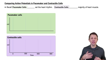
Comparing Action Potentials in Pacemaker and Contractile Cells

Contractile Cardiac Muscle Cells

Contractile cardiac muscle cells, found throughout the heart, are responsible for the actual contraction and pumping of blood. These cells respond to the electrical signals generated by pacemaker cells, leading to coordinated contractions. They contain myofibrils and are structured to facilitate strong contractions, ensuring effective blood circulation.
Recommended video:
Guided course
6:36
Comparing Action Potentials in Pacemaker and Contractile Cells

Autorhythmicity

Autorhythmicity refers to the ability of certain cardiac cells, particularly pacemaker cells, to generate rhythmic electrical impulses without external stimulation. This intrinsic property is crucial for maintaining a consistent heart rate and rhythm, allowing the heart to function autonomously and adapt to the body's varying demands for blood flow.
Recommended video:
Guided course
8:47
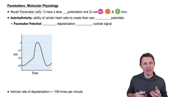
Pacemakers: Molecular Physiology