Ch. 13 The Peripheral Nervous System
Amerman - Human Anatomy & Physiology 2nd Edition
Erin C. Amerman2nd EditionHuman Anatomy & PhysiologyISBN: 9780136873822Not the one you use?Change textbook
Chapter 13, Problem 14

The lower motor neurons that innervate contractile skeletal muscle fibers are called:
a. α-motor neurons
b. β-motor neurons
c. upper motor neurons
d. γ-motor neurons

Verified step by step guidance
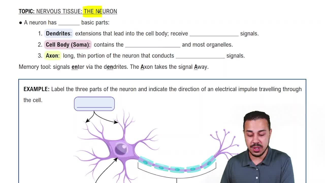
1
Understand the role of motor neurons: Motor neurons are responsible for transmitting signals from the central nervous system to muscles, causing them to contract. They are divided into upper motor neurons and lower motor neurons.
Differentiate between upper and lower motor neurons: Upper motor neurons originate in the brain and send signals to lower motor neurons. Lower motor neurons directly innervate skeletal muscle fibers to cause contraction.
Learn about the types of lower motor neurons: Lower motor neurons are further classified into α-motor neurons, β-motor neurons, and γ-motor neurons, each with specific functions.
Focus on α-motor neurons: α-motor neurons specifically innervate extrafusal muscle fibers, which are the contractile fibers of skeletal muscles responsible for generating force.
Eliminate incorrect options: β-motor neurons innervate intrafusal fibers and are involved in muscle spindle sensitivity. γ-motor neurons also innervate intrafusal fibers to adjust spindle tension. Upper motor neurons do not directly innervate skeletal muscle fibers. Therefore, the correct answer is α-motor neurons.

Verified video answer for a similar problem:

This video solution was recommended by our tutors as helpful for the problem above.
Was this helpful?

Key Concepts

Here are the essential concepts you must grasp in order to answer the question correctly.

Lower Motor Neurons

Lower motor neurons are the neurons that directly innervate skeletal muscle fibers, facilitating muscle contraction. They originate in the spinal cord and extend to the muscles, transmitting signals that lead to voluntary movements. Understanding their role is crucial for identifying how motor commands are executed in the body.
Recommended video:

α-Motor Neurons

α-motor neurons are a specific type of lower motor neuron responsible for innervating extrafusal muscle fibers, which are the primary contractile units of skeletal muscles. They play a vital role in generating force and movement by transmitting action potentials that lead to muscle contraction. Recognizing their function is essential for answering questions about muscle innervation.
Recommended video:

Motor Neuron Types

Motor neurons can be categorized into different types, including α-motor neurons, β-motor neurons, and γ-motor neurons. Each type has distinct functions; for instance, γ-motor neurons innervate intrafusal fibers in muscle spindles, while β-motor neurons are involved in modulating muscle tone. Understanding these distinctions is important for comprehending the overall motor control system.
Recommended video:
Related Practice
Textbook Question

Which parts of the body have the greatest amount of space dedicated to them in the primary somatosensory cortex? Why?

370
views
Textbook Question

List and describe the basic steps involved in producing movement, beginning with the upper motor neurons in the cerebral cortex.

382
views
Textbook Question

Which of the following statements is false?

a. The spinothalamic tracts are part of the anterolateral system.

b. Pain, temperature, and crude touch stimuli are carried by the anterolateral system.

c. Descending pathways from the brain and spinal cord can make the spinal cord less receptive to pain stimuli.

d. The thalamus serves as the 'gateway' for entry of all special sensory stimuli into the cerebral cortex, with the exception of audition (hearing).

394
views
Textbook Question

The cell bodies of upper motor neurons reside in the ________ and function to ________, whereas the cell bodies of lower motor neurons reside in the ________ and function to ________.

515
views
Textbook Question

______detect the degree to which a muscle is stretched, whereas______detect the force of a muscle contraction.

351
views
Textbook Question

Label the following components of the corticospinal tracts with numbers 1 through 6, with 1 being the origin of the tracts and 6 their destination.     

_____ Medullary pyramids where most fibers decussate.     

_____ Anterior horn of the spinal gray matter.     

_____ Midbrain and pons.      

_____ Upper motor neurons in the primary motor and premotor cortices.     

_____ Corona radiata and internal capsule.        

_____ Lateral funiculus of the spinal cord.

395
views