Textbook Question
What aspect of DNA structure makes it possible for the proteins of nucleotide excision repair to recognize many different types of DNA damage?
(c) the energy differences between correct and incorrect base pairs
1427
views

 Verified step by step guidance
 03:37
03:37 02:37
02:37 03:56
03:56What aspect of DNA structure makes it possible for the proteins of nucleotide excision repair to recognize many different types of DNA damage?
(c) the energy differences between correct and incorrect base pairs
What aspect of DNA structure makes it possible for the proteins of nucleotide excision repair to recognize many different types of DNA damage?
(d) the regularity of DNA's structure
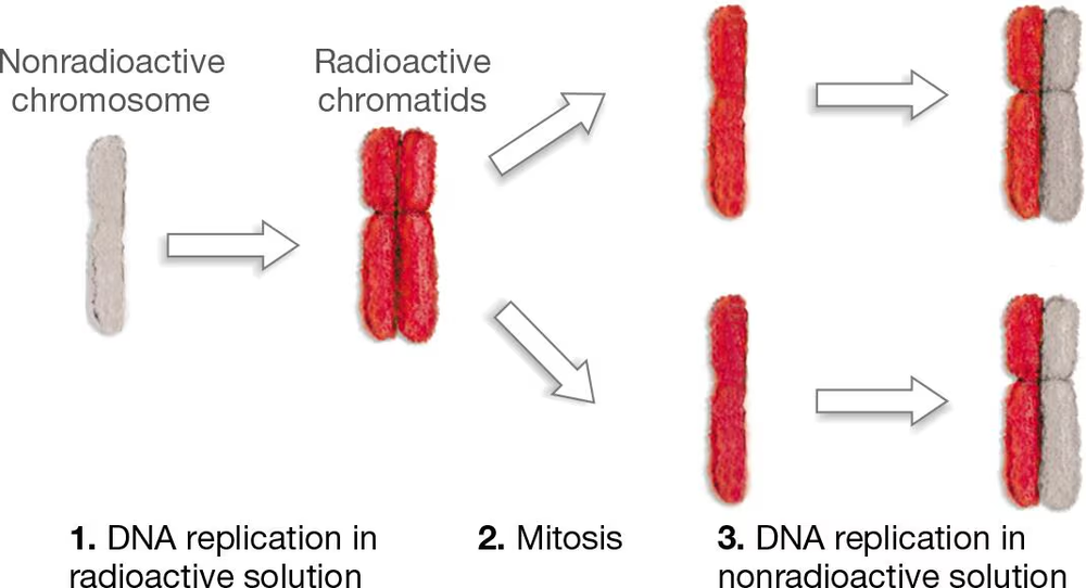
In the late 1950s, Herbert Taylor grew bean root-tip cells in a solution of radioactive thymidine (a precursor to one of the deoxyribonucleotides in DNA) and allowed them to undergo one round of DNA replication. He then transferred the cells to a solution without radioactive thymidine, allowed them to replicate again, and examined their chromosomes for the presence of radioactivity. His results are shown in the following figure, where red indicates a radioactive chromatid.
(b) What would the results of Taylor's experiment be if eukaryotes used a conservative mode of DNA replication?
The graph that follows shows the survival of four different E. coli strains after exposure to increasing doses of ultraviolet light. The wild-type strain is normal, but the other strains have a mutation in either a gene called uvrA, a gene called recA, or both.
(a) Which strains are most sensitive to UV light? Which strains are least sensitive?
The graph that follows shows the survival of four different E. coli strains after exposure to increasing doses of ultraviolet light. The wild-type strain is normal, but the other strains have a mutation in either a gene called uvrA, a gene called recA, or both.
(b) What are the relative contributions of these genes to the repair of UV damage?