Pearson+ LogoPearson+ Logo
Ch. 16 - Adaptive Immunity
Bauman - Microbiology with Diseases by Taxonomy 6th Edition
Bauman6th EditionMicrobiology with Diseases by TaxonomyISBN: 9780134832302Not the one you use?Change textbook
Chapter 16, Problem 2

The nearby image is a transmission electron micrograph of a dendritic cell. Indicate where a scientist could find molecules of MHCI and MHC II. Label a pseudopod and a vesicle.
<IMAGE>

Verified step by step guidance
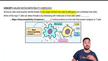
1
Step 1: Identify the pseudopods on the dendritic cell. Pseudopods are the finger-like projections extending from the cell membrane, used by the dendritic cell to interact with its environment and capture antigens.
Step 2: Locate vesicles inside the dendritic cell. Vesicles appear as small, membrane-bound sacs within the cytoplasm and are involved in processing and transporting molecules such as antigens and MHC complexes.
Step 3: Understand where MHCI molecules are found. MHCI molecules are expressed on the surface of almost all nucleated cells, including dendritic cells, so they will be present on the plasma membrane, including the pseudopods.
Step 4: Understand where MHCII molecules are found. MHCII molecules are primarily expressed on professional antigen-presenting cells like dendritic cells and are found both on the plasma membrane and within intracellular vesicles where antigen processing occurs.
Step 5: Label the pseudopods as the membrane extensions, the vesicles as the internal membrane-bound structures, and indicate that MHCI molecules are on the cell surface (including pseudopods), while MHCII molecules are both on the surface and inside vesicles involved in antigen processing.

Verified video answer for a similar problem:

This video solution was recommended by our tutors as helpful for the problem above.
Video duration:
2m
Was this helpful?

Key Concepts

Here are the essential concepts you must grasp in order to answer the question correctly.

Major Histocompatibility Complex Class I (MHCI)

MHCI molecules are found on the surface of all nucleated cells, including dendritic cells. They present endogenous antigens, typically from intracellular pathogens, to CD8+ cytotoxic T cells. In the electron micrograph, MHCI molecules would be located on the plasma membrane of the dendritic cell, including the pseudopods.
Recommended video:
Guided course
02:31
Major Histocompatibility Complex Molecules

Major Histocompatibility Complex Class II (MHCII)

MHCII molecules are primarily expressed on professional antigen-presenting cells like dendritic cells. They present exogenous antigens, derived from extracellular pathogens, to CD4+ helper T cells. MHCII molecules are found on the plasma membrane and within intracellular vesicles involved in antigen processing.
Recommended video:
Guided course
02:31
Major Histocompatibility Complex Molecules

Pseudopods and Vesicles in Dendritic Cells

Pseudopods are membrane extensions used by dendritic cells for movement and antigen capture, visible as finger-like projections in the micrograph. Vesicles are membrane-bound compartments inside the cell that transport and process antigens; they appear as small, round structures within the cytoplasm. Both structures are crucial for antigen uptake and presentation.
Recommended video:
Guided course
05:20
Cell Inclusions