Textbook Question
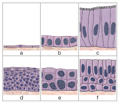
Identify the six types of epithelial tissue shown in the drawing below.
c. ___
178
views

 Verified step by step guidance
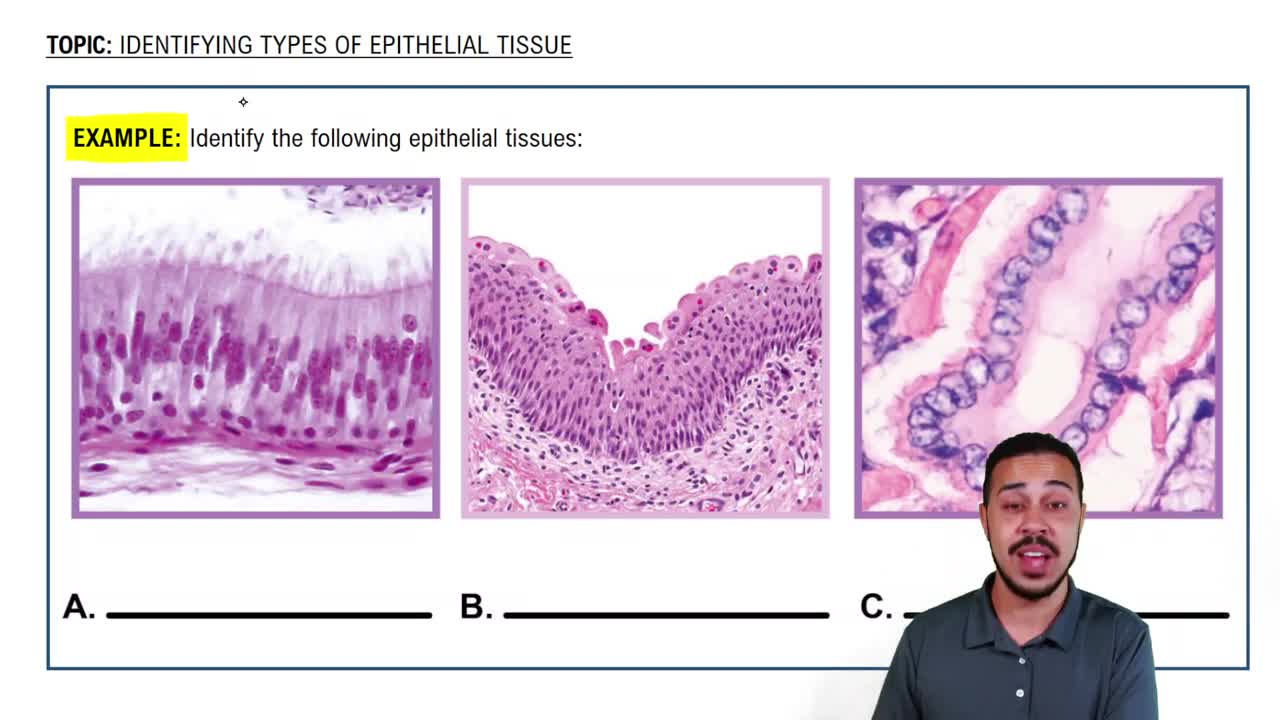
 07:25
07:25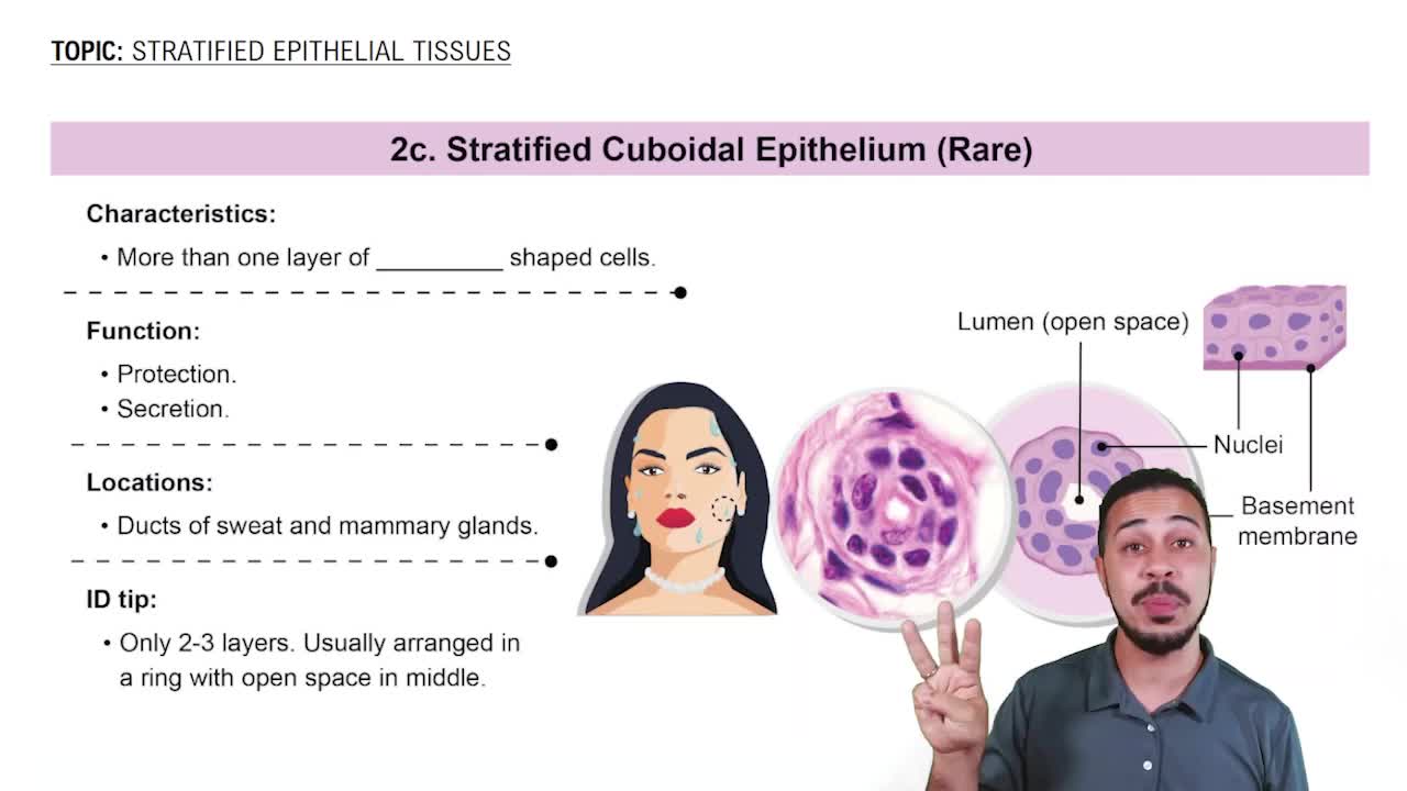 08:05
08:05 03:54
03:54Identify the six types of epithelial tissue shown in the drawing below.
c. ___
Identify the six types of epithelial tissue shown in the drawing below.
d. ___
Identify the six types of epithelial tissue shown in the drawing below.
b. ___