Identifying epithelial tissue is essential in understanding its structure and function within the body. Epithelial tissues are characterized by tightly packed cells that form a protective barrier, with one side adjacent to an open space. To effectively identify different types of epithelial tissue, a systematic approach using a flowchart can be beneficial.
The identification process begins by examining a micrograph of epithelial tissue. The first question to consider is whether the cells are tightly packed with one side adjacent to an open space. If the answer is yes, the tissue is likely epithelial. If no, it may be another type of tissue.
Next, determine if there are two or more clearly defined layers of cells. If there are multiple layers, the tissue is classified as stratified epithelial tissue. If not, it is classified as simple epithelial tissue. Simple epithelial tissues can be further categorized based on cell shape:
1. **Simple Squamous Epithelium**: If the cells appear flat or ribbon-like, they are classified as squamous. This type is typically found in areas requiring rapid diffusion, such as the air sacs of the lungs and capillaries.
2. **Simple Cuboidal Epithelium**: If the cells are cube-shaped, they are classified as cuboidal. This type is often found in gland ducts and kidney tubules.
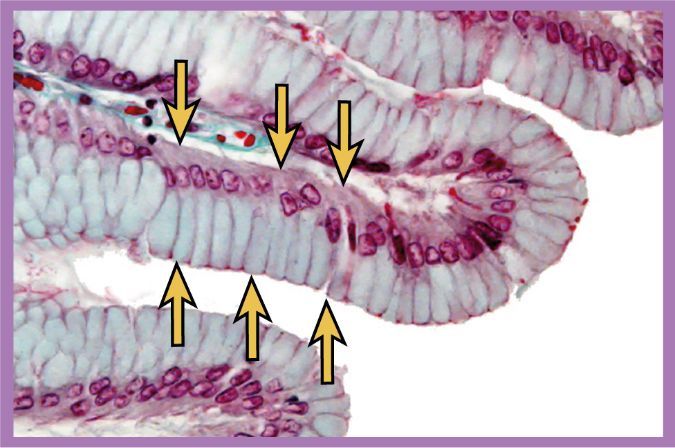
3. **Simple Columnar Epithelium**: If the cells are tall and narrow, they are classified as columnar. This type is commonly found in the digestive tract and may have microvilli for absorption or cilia for movement of substances.
In cases where the nuclei of columnar cells are aligned in a single row, the tissue is identified as simple columnar. If the nuclei are at different levels, it indicates pseudostratified columnar epithelium, which appears stratified but is not, as all cells contact the basement membrane. This type is often found in the respiratory tract and may contain goblet cells that secrete mucus.
For stratified epithelial tissues, if there are many layers, the next step is to determine the shape of the surface cells:
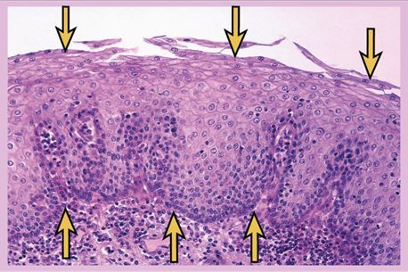
1. **Stratified Squamous Epithelium**: If the surface cells are flat and may flake off, the tissue is stratified squamous. This type can be keratinized (found in the skin) or unkeratinized (found in moist areas like the mouth).
2. **Transitional Epithelium**: If the surface cells are pillow-shaped, the tissue is transitional, which is unique to the urinary system and allows for stretching.
If there are only 2 to 3 layers of cells, the tissue can be either stratified columnar or stratified cuboidal. Stratified columnar epithelium has tall, narrow surface cells and is found in areas where different epithelial types meet. Stratified cuboidal epithelium has cube-shaped surface cells and provides more protection, typically found in larger ducts of glands.
Understanding these classifications and characteristics of epithelial tissues is crucial for identifying them in histological studies and appreciating their roles in various physiological processes.