Before a cell can divide, it must first replicate its DNA, which is crucial for ensuring that each daughter cell receives an accurate copy of the genetic material. The organization of DNA within the cell plays a significant role in this process. The complete set of all a cell's DNA is referred to as the genome, which encompasses all the genetic material that determines the inherited traits of an organism, primarily consisting of DNA.
Within the cell, DNA associates with proteins known as histones. These histones form structures called nucleosomes, which consist of a core of eight histone proteins around which DNA is wrapped. This organization can be visualized as yarn wrapped around a pencil, where the pencil represents the histone core and the yarn symbolizes the DNA.
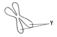
DNA organization varies depending on the cell's state. In a non-dividing cell, the DNA is loosely packed into a structure called chromatin. Chromatin allows for easier access to the genetic material for processes such as transcription and replication. In contrast, when a cell prepares to divide, the nucleosomes condense into tightly packed structures known as chromosomes. This condensation is essential for the proper segregation of DNA during cell division, ensuring that each new cell receives the correct amount of genetic material.
To summarize, chromatin represents the loosely coiled form of DNA found in non-dividing cells, while chromosomes are the tightly packed form of DNA present in dividing cells. Understanding this organization is vital for grasping how genetic information is managed and transmitted during cell division.