Simple epithelial tissues are a fundamental category of epithelial tissues characterized by a single layer of cells that directly contact the basement membrane. In human anatomy, there are eight types of epithelial tissues, with four classified as simple epithelial tissues. The term "simple" signifies that these tissues consist of only one layer, distinguishing them from stratified epithelial tissues, which will be discussed later.
Understanding simple epithelial tissues is crucial as they play vital roles in various physiological functions, including absorption, secretion, and filtration. The four types of simple epithelial tissues include:
- Simple Squamous Epithelium: This type consists of flat, scale-like cells and is involved in processes such as diffusion and filtration.
- Simple Cuboidal Epithelium: Comprising cube-shaped cells, this tissue is primarily involved in secretion and absorption.
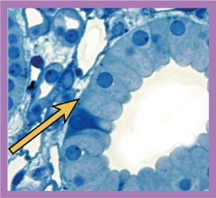
- Simple Columnar Epithelium: Featuring tall, column-like cells, this type is often found in areas where absorption and secretion occur, such as the digestive tract.
- Pseudostratified Columnar Epithelium: Although it appears stratified, this tissue is a single layer with varying cell heights, commonly found in the respiratory tract.
Each type of simple epithelial tissue has unique structural characteristics and functions, which will be explored in detail in subsequent lessons. Understanding these tissues is essential for grasping their roles in the human body and their implications in health and disease.