Textbook Question
List the similarities and differences among the three types of muscle tissue.
76
views

 Verified step by step guidance
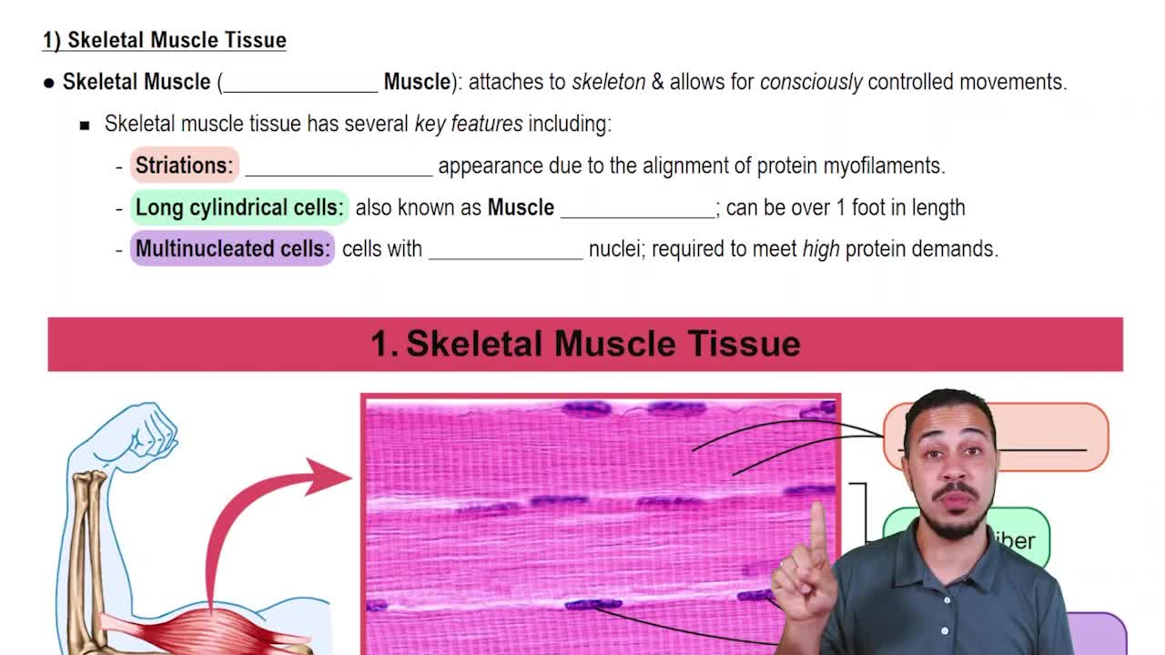
 06:56
06:56 06:29
06:29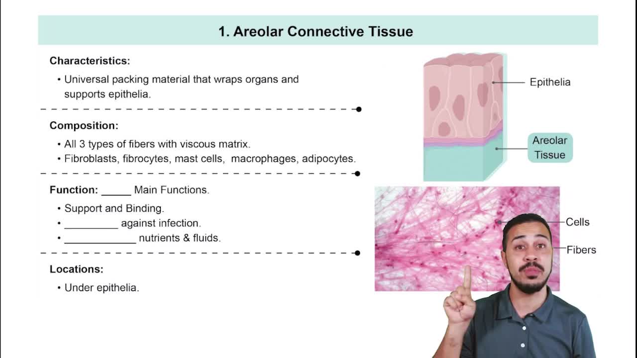 08:25
08:25List the similarities and differences among the three types of muscle tissue.
A layer of glycoproteins and a network of fine protein filaments that prevents the movement of proteins and other large molecules from the connective tissue to the epithelium describe
(a) Interfacial canals
(b) The basement membrane
(c) The reticular lamina
(d) Areolar tissue
(e) Squamous epithelium
Why does damaged cartilage heal slowly?
(a) Chondrocytes cannot be replaced if killed, and other cell types must take their place.
(b) Cartilage is avascular, so nutrients and other molecules must diffuse to the site of injury.
(c) Damaged cartilage becomes calcified, thus blocking the movement of materials required for healing.
(d) Chondrocytes divide more slowly than other cell types, delaying the healing process.
(e) Damaged collagen cannot be quickly replaced, thereby slowing the healing process.