What evidence suggests that during anaphase, kinetochore microtubules shorten at the kinetochore?
Verified step by step guidance
1
1. Anaphase is a stage in mitosis and meiosis where the chromosomes separate. Each chromosome is attached to a structure called a kinetochore, which is connected to the spindle fibers (microtubules).
2. The evidence that suggests kinetochore microtubules shorten at the kinetochore during anaphase comes from several observations. One of these is the movement of chromosomes towards the poles of the cell during anaphase. This movement is consistent with the shortening of the microtubules at the kinetochore end.
3. Another piece of evidence comes from experiments where fluorescent markers are used to label the microtubules. When observed under a microscope, these markers appear to move towards the poles of the cell during anaphase, suggesting that the microtubules are shortening from the kinetochore end.
4. Additionally, electron microscopy has shown that during anaphase, the kinetochore appears to remain attached to the end of the microtubule, even as the chromosome moves towards the pole. This suggests that the microtubule is shortening from the kinetochore end, pulling the chromosome along with it.
5. Finally, biochemical studies have shown that the protein complexes that form the kinetochore can catalyze the disassembly of microtubules, providing a mechanism for how the microtubules could be shortened at the kinetochore end during anaphase.
Verified video answer for a similar problem:
This video solution was recommended by our tutors as helpful for the problem above
Video duration:
2m
Play a video:
0 Comments
Key Concepts
Here are the essential concepts you must grasp in order to answer the question correctly.
Anaphase
Anaphase is a stage in mitosis where sister chromatids are pulled apart toward opposite poles of the cell. This process is crucial for ensuring that each daughter cell receives an identical set of chromosomes. During anaphase, the cohesin proteins that hold the sister chromatids together are cleaved, allowing them to separate and move due to the action of the spindle apparatus.
Kinetochore microtubules are specialized structures that connect the spindle apparatus to the kinetochore, a protein complex assembled on the centromere of each chromosome. These microtubules play a vital role in chromosome movement during cell division by exerting forces that pull the chromatids apart. Their dynamic nature allows them to grow and shrink, facilitating the separation of chromosomes.
Microtubule shortening refers to the process where the length of microtubules decreases, which is essential during anaphase for the movement of chromosomes. This shortening occurs through the depolymerization of tubulin subunits at the kinetochore end of the microtubules. Evidence for this process can be observed through live-cell imaging and experiments that track the dynamics of microtubules during cell division.

 Verified step by step guidance
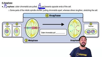
 02:39
02:39