Join thousands of students who trust us to help them ace their exams!
Multiple Choice
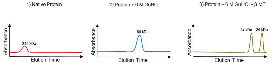
A new protein of unknown structure has been purified & gel filtration chromatography reveals that the native protein has a molecular weight of 240 kDa.Chromatography in the presence of 6 M guanidine hydrochloride (GuHCl), a chaotropic agent that has a similar effect on proteins as urea, yields a single absorbance peak corresponding to a protein of Mr 60 kDa. Chromatography in the presence both of 6 M guanidine hydrochloride and 10 mM β-mercaptoethanol (β-ME) yields peaks for proteins of Mr 34 kDa and 26 kDa. Using this data, which option best describes the structure of this protein? Hint: sketch a visual of the protein after each chemical treatment.
A
A homotetramer (4 identical 60 kDa subunits); each subunit is a heterodimer of 2 disulfide-linked chains (34 & 26 kDa).
B
A heterooctomer (8 different subunits); four subunits each of 34-kDa & 26 kDa, all held together via disulfide bonds.
C
A homodimer (2 identical 120 kDa subunits); each subunit is a a homodimer of 2 disulfide-linked chains (60 kDa each).
D
A heterotetramer (4 different subunits); each subunit is a homodimer of 2 disulfide-linked chains (60 kDa each).
0 Comments
Verified step by step guidance
1
Begin by analyzing the native protein's molecular weight, which is 240 kDa, as shown in the first chromatogram. This suggests that the protein is a multimeric complex.
Next, consider the effect of 6 M guanidine hydrochloride (GuHCl), a chaotropic agent that denatures proteins by disrupting non-covalent interactions. The second chromatogram shows a single peak at 60 kDa, indicating that the native protein dissociates into smaller subunits under denaturing conditions.
Now, examine the effect of both 6 M GuHCl and 10 mM β-mercaptoethanol (β-ME), which reduces disulfide bonds. The third chromatogram reveals two peaks at 34 kDa and 26 kDa, suggesting that each 60 kDa subunit is composed of two smaller chains linked by disulfide bonds.
Sketch a visual representation of the protein structure after each chemical treatment: initially as a 240 kDa complex, then as four 60 kDa subunits after GuHCl treatment, and finally as eight chains (four 34 kDa and four 26 kDa) after both GuHCl and β-ME treatment.
Based on the data, conclude that the protein is a homotetramer composed of four identical 60 kDa subunits, each subunit being a heterodimer of two disulfide-linked chains (34 kDa and 26 kDa).


 Verified step by step guidance
