So now that we've introduced tertiary structure, we can talk about protein motifs and domains. So it turns out that tertiary protein structure actually includes the distribution of secondary structures such as Alfa Ulysses, beta strands, beta turns and loops to make protein motifs and protein domains. And so, just to get you guys oriented on the hierarchy of all of these terms and structures, we've got this image below. And so, of course, at the lowest level of the hierarchy, What we have is our primary protein structure. And our primary protein structure is just the combination of amino acid composition as well as amino acid sequence and the sequences the particular order of amino acids from the N terminal end to the C terminal end of a poly peptide chain. So then what we have is our secondary structures, and we talked about multiple types of secondary structures, including Alfa Helix sees beta sheets and loops and turned loops and beta turns. And so, uh, then what we did was we jumped straight from secondary structure to tertiary structure, and now that we've got tertiary structure introduced, we can better understand this organization. So it turns out that secondary structures actually come together to form super secondary structures, which are also known as protein motifs. And then the super secondary structures can be organized into particular domains. And then the domains are just independent folding units. And so, together, the motifs in the domains. As you can see by this bracket here, uh, they come together to form the tertiary structure, which we know as the overall three D shape off the protein. And so, uh, what we're going to first do is talk about the super secondary structures or the motifs. And then once we're done talking about the motifs, we'll go and we'll talk about the protein domains. So I'll see you guys in our next video or we'll talk about protein motifs.
2
concept
Protein Motifs and Domains
3m
Play a video:
Was this helpful?
So, as we said in our previous video, motifs are also known as super secondary structures. And if you Google, the definition of motif, which will find is that in the everyday language, it means a pattern. And that's really all a protein motif is. It's literally just a specific pattern and combination of secondary structures such as, for instance, Alfa Ulysses, beta strands, beta turns and loops. And so motifs can have varying functions or varying jobs in different proteins, and so they can have different functions and proteins that include providing strength as well as creating binding sites for different molecules. And so it turns out that there are actually several hundreds of known motifs, and so it's not likely that your professor is gonna want you to memorize all of them. So, uh, there are a few common ones that you might wanna know, and so I've tried to provide those here. And so what you'll see is that we've got a helix loop helix motif, a coiled coil motif, beta alfa beta motif and a hairpin motif. And so, in our example below, what we're going to do is label the corresponding motifs, and so for our first motif here. What we have is the helix loop helix motif and literally this is just an Alfa helix. So notice it's an Alfa helix with a loop and another Alfa helix. And that's exactly what the helix loop helix motif is so pretty easy. Now, the next one that we have is the coiled coil, and so the coiled coil. What you'll see is that it's, uh, Alfa Helix is when you have to Alfa. He leases that are actually wind it up on one another. And so remember that a single Alfa helix is going to be an interchange structure. So it's gonna be within one single poly peptide chain and so on. Alfa Helix is different than a coiled coil because the Alfa helix is a single of poly peptide chain, whereas the coiled coil is to poly peptide chains to Alfa Helix structures. That air wind it up and twisted on one another. And this type of structure really does create a lot of strength. So whenever you see coiled coil structures, you know that that protein is gonna provide a lot of strength. So the next motif that we have is the beta Alfa beta motif. And so, with this beta alfa beta motif, it's literally just a beta strand connected to an Alfa helix, which is connected to another beta strand. So it's beta alfa beta, pretty straightforward. So then our last and final motif is the hairpin motif, and with the hairpin motif, it's literally just to beta strands going on anti parallel direction that's looped together by a hairpin loop. So it's a small little hairpin loop that connects to beta strands that are anti parallel. They're going in opposite directions. And so these were some of the common motifs that you might want to try to commit to memory that your professor might try to ask you guys about. And so we'll be able to get a little bit of practice in our practice video, so I'll see you guys there
3
Problem
Which of the following colored regions in the images below is not an example of a supersecondary structure?
A
Greek key.
B
β-meander.
C
βαβ unit.
D
Type I β-turn
E
Helix-loop-helix
4
concept
Protein Motifs and Domains
3m
Play a video:
Was this helpful?
So now that we've talked about protein motifs, we can focus on protein domains, and so domains are really just combinations of motifs. And so again, motifs are just patterns and combinations of secondary structures and domains are combinations of motifs, so you can see how that hierarchy builds up on itself. And really, the interesting thing about domains is that they are independently folding units, so they have the ability to independently fold from the rest of the protein. And also, domains have the potential toe have their own discreet functions. And so that's quite interesting. And so another thing that you should know about domains is that they are literally an extension to the peptides backbone, and so domains can be found on a single poly peptide chain. One single poly peptide chain could have multiple domains because literally a domain is just the an extension to the peptide back mode. So it's really important not to get domains confused with sub units. And so sub units over here is really a term that we're going to talk more about when we get to Quaternary structure a little later in our course. But sub units are completely separate poly peptide chains and their backbones are not connected. But again, domains are literally just an extension to the peptides backbone. And so they are very different terms, and it's important to point that out now just to make sure that you're starting to get that, make that realization. And so again, we'll revisit this term sub units when we get to question a restructure later in our course now, another very important thing to notice about domains is that they're often grouped according to the structural and functional characteristics that they have, and so scientists can actually learn Ah, lot about, um, they can learn a lot from studying domains. So say a scientists find a brand new protein that they know nothing about. And within this brand new protein, ah, scientists recognizes a smaller particular domain within that big protein and that domain that they recognize. They've studied that domain in another protein, and they learned a lot about that domain. And so, by knowing this particular domain, that's in this new protein that can reveal some of the functions that this new protein has. And so that's why it's very important and critical to study a proteins domains. And so, in our example below what we're gonna do is circle the three most obvious domains in the figure. And so in this figure, down below, this is literally one single poly peptide chain that is completely folded up. And so we've colored the different domains that make it pretty easy to see. So what you'll see is that we've got one domain in the back. Over here in purple. We've got another domain in the middle here in green, and then we've got our last domain over here in blue. And so what you can see is that even though this is a single poly peptide chain, it has these independently folding units. And so if you were to cut off, say this, uh, purple section over here from the rest of the chain, this, uh, section because it's it's own domain, we would expect it to still fold properly. And the same goes for all of these other domains here. So thes air the three domains. And so, uh, this concludes our lesson on protein motifs and domains and will be able to get some practice in our practice video. So I'll see you guys there
5
Problem
Which of the following statements concerning a Cas9 endonuclease’s protein domains is true?
A
They are a form of its secondary structure.
B
They have only been found in eukaryotic proteins.
C
All its domains consist of separate polypeptide subunits.
D
They retain their shape when separated from the protein.
6
Problem
Which of the following is true concerning the motifs and domains of proteins?
A
Many domains make up a motif.
B
Every polypeptide chain is limited to one domain.
C
Separate proteins with similar domains are likely to have a similar function.
D
All domains of a protein have the same function.
7
Problem
Appropriately label the domains of the membrane embedded protein in the figure below.
Was this helpful?
8
Problem
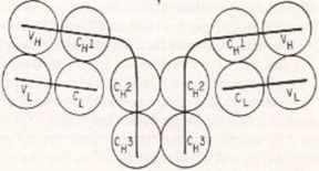
The structure of an immunoglobulin G (antibody) molecule is shown schematically below. The black solid lines depict individual polypeptides and so there are four polypeptides in the quaternary structure of this molecule. Each of the spheres represents a stretch of about 100 amino acids folded independently of the rest of the polypeptide and performs a specific function in the molecule. Therefore, each sphere was given its own individual name (VH, CH1, CH2, etc.). Without knowing any additional details, you can predict that there must be TWELVE ___________________ in this molecule.
A
α-helices.
B
Domains.
C
Subunits.
D
Motifs.
9
Problem
What is the main difference between an endonuclease’s domains and its subunits?
A
Its domains are composed mostly of the α-helix, while subunits contain both α-helices and β-sheets.
B
Its subunits are separate polypeptide chains, while its domains constitute a part of a polypeptide chain.
C
Its domains do not have secondary structure, whereas its subunits have do.
D
Its domains are stabilized by hydrogen bonds, while its subunits are stabilized by disulfide bonds.