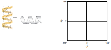
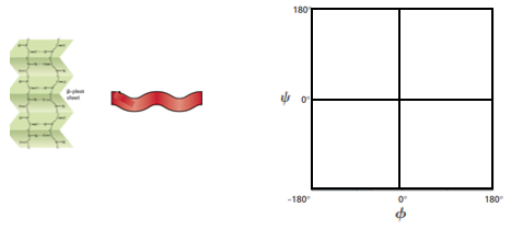
in this video, we're gonna begin our discussion on Rama Condron plots. So in our last lesson video, we said that peptide bond rotation is limited and restricted due to its partial double bond character. But bond rotations specifically around the Alfa Carbon bonds are still possible, even though the peptide bond rotation is restricted. And it turns out that there are three bond rotation angles that you guys should be familiar with, and they're symbolized with these Greek letters and so down below. In our example, we're gonna distinguish between these three bond angles in this peptide that's being shown. And so in this peptide we have a free amino group on the far left and on the far right. What we have is a little squiggly line which represents a continuation of the peptide. So we're not being shown the C terminal end and we're on Lee being shown a small little snippet of our peptide, and so notice that the are groups of our peptide here are in blue. And so the first amino acid residue has a hydrogen as its our group, which means that we have a glazing amino acid residue and the second amino acid residue has a metal group, as it's our group, which means that we have an Alan een amino acid residue. And so to distinguish between these three bond angles, the first bond angle that you guys should know is the five bond angle. And FYI is symbolized with the Greek letter Phi, and it represents the rotation angles around the Alfa carbon nitrogen bond in the backbone. And so, taking a look at our Allan, an amino acid residue down below in our peptide noticed that the Alfa Carbon is this carbon that's here, which is attached to the our group. And so the Alfa Carbon bonded to the nitrogen is going to represent the fi bond. And so you can see that this bond right here is the fi Bond and Fi represents the bond rotation angles around the fi bond, so the second bond rotation angle that you guys should know is the sigh Bond and Sigh is symbolized with the Greek letter sigh, and it represents rotation angles around the Alfa Carbon Carbonnel group carbon bond in the backbone. And so, taking a look at our Allen and amino acid residue again, here is our Alfa Carbon and the Carbonnel Group. Carbon is over here. And so the bond between the Alfa Carbon and the Carbonnel Group Carbon is the side bond and Sigh represents the bond rotation angles around the side bond. And so the last bond rotation angle that you guys should be familiar with is Omega, and Omega is symbolized with the Greek letter omega, and it represents rotation angle of the peptide bond. And so, looking below at our peptide, we can see that the peptide bond is in red over here and notice that it has partial double bond character with this dotted line. And Omega is shown here to represent the bond rotation angles around the peptide bond. Now we know that Omega and the bond rotation angles around the peptide bond is severely limited and restricted because of the partial double bond character. And because it's restricted, it's not really interesting toe plot Omega on a Rama Condron plot. And so it turns out that Omega is actually excluded from a Rama Condra plot, and when we're plotting a Rama Condron plot, we only focus on the fi and the Sai bond rotation angles. And so, looking over here, at our other figure. What we have is a ball and stick figure of a peptide and noticed that the Alfa Carbon is this black ball that's here in the center. And so the bonds that are around the Alfa Carbon are all able to still rotates. So bond rotations around the Alfa Carbon are still possible. And so that's why you see that the Alfa Carbon nitrogen bonds of this blue ball over here represents the nitrogen. We said that the Alfa Carbon nitrogen bond is the fi bond. So this bond here in green is analogous to this bond over here in green, which is the five bond. And then the side bond is going to be this bond over here and yellow between the Alfa Carbon and the Carbonnel group carbon. And so you can see that these arrows that are being shown are indicating that these bonds are able to freely rotate, whereas the peptide bond, which would be, uh, this bond over here in red and this bond over here, these bonds are restricted and limited, and that's why we see the plane behind them that indicates the uh, peptide group that is plainer and So what we what this means because we're mainly gonna be focusing on fi and Side bonds is that we need a way to distinguish between the fire and the side bonds. And so what helps me is that when you actually write out five, um, you'll see that the H here really looks like a larger version of a lower case end. And the end here reminds you of the nitrogen atom. And so that can tell you that thief I Bond is really just the Alfa Carbon nitrogen bond. And that's exactly what Fi is all about. It's the Alfa Carbon nitrogen bond. And again, both the fi and the SAI have the Alfa Carbon. So really distinguishes them is the nitrogen and the carbonnel group carbon. And so, if you can remember that this little H here looks like a lower case N, which reminds you of the nitrogen atom. Then you'll remember that fi as the Alfa Carbon nitrogen bond Now for Cy. On the other hand, when you draw the Greek letter side, you literally draw sideways, see with a line through it, and so the sideways see can remind you of a carbon and So the side bond is all about the Alfa Carbon carbon bonds. So there's Onley carbons that are involved, and so the sigh here can remind you of the side bond. And so, if you can remember these memory tools, you'll be able to distinguish between the fi and the Sai bonds. And so the last thing I want to leave you guys off with is that within a peptide, each individual amino acid residue has its own fi and side bonds. And so if we take a look at our glazing amino acid residue over here, notice that it's Alfa Carbon is right here. And so the Alfa Carbon nitrogen bond, which would be this bond right here, is going to be the fi bond. Just like we said before, Alfa Carbon Nitrogen is the five bond. So what we can do is we can draw on our Greek symbol, Fi right there. And so the Alfa Carbon carbon bond over here is going to be the Cy Bond. And so this bond here in yellow is gonna be the Cy Bond so we can draw in our Greek symbol side. And then, of course, the peptide bonds are always going to be. So this pond right here is going to be the peptide bond, and that's gonna represent Omega, which we can draw in here as well. And so moving forward, we're gonna talk, Maura about Rama Condron plots, and we'll be able to get, ah, lot more practice understanding Fi inside bond angles after we talk about Rama condom plots. And so I'll see you guys in our next video, where we'll be able to get a little bit of practice distinguishing between fi and side bond angles, so I'll see you guys there.
2
concept
Ramachandran Plot
6m
Play a video:
Was this helpful?
So now that we've talked about the fi sigh in Omega Bond angles, we can talk about Rama Condron plots. And since the peptide bond rotation or Omega is hindered by the double bond nature, really, it's just the fi and the Sai Bond angles that determined the structure of a poly peptide. And so because it's fi inside that determine the structure of a poly peptide, that is what the Rama Condron plot focuses on plotting. And so the Omega Bond angle is actually excluded from the Rama Condron plot. So it's not part of the Rama Condron plot. And so even though the fi in the side bond angles are free and they are free to rotate, they're still a little bit restricted. They're still somewhat restricted, and they're restricted by Starik. Hindrance specifically, between the are groups and the backbone, Carbonnel Oxygen's. And so the rotation angles for five and Sigh actually ranged from negative 182 0 or counterclockwise angles, as well as from zero to positive 180 which would be clockwise angles. And so when we're talking about a Rama Condron plot, really, what a Rama condom plot is showing is just the permissible and the non permissible bond angles for fi and Sigh. And, um, when you think about a Rama condom plot, you can think of it as showing just the fine side angles for a single amino acid residue. Or you can think of it as showing all of the find sai angles for all of the amino acid residues in an entire protein. And so, in our example below, What we're gonna do is fill in the blanks on the Rama Condron plot, and so notice that this here is actually a typical Rama Condron plot for pretty much all of the amino acids. Except a few exceptions, except just two exceptions, which we'll talk about later. But all of the Rama condom plots pretty much look like this one that's shown here. And so what you'll notice is that on the X axis, what's being plotted are the fi bond angles, and so what we can do is in this blank. We can put the five bond angles and notice that the five bond angles range from negative 182 positive 180 degrees, and then on the Y axis What we have is the sigh bond angles, and we can help you remember that the side bond angles goes on. The Y axis is that the sigh bond angles go on the side of the Rama Condron plot and so notice that the side bond angles also ranged from negative 180 upto positive 180 degrees. And so again, all aroma Condron plot is showing are the permissible and non permissible angles. And so what you'll see is that we have these blue regions, and then we have the white regions, and so the blue regions represent permissible bond angles. And so the dark blue regions represent mawr. Permissible bond angles and the light blue regions represent less permissible bond angles. And then, of course, the white regions represent non permissible bond angles. So that means that these bond angles cannot be achieved because of Stare Kendricks between either the are groups or the Carbonnel group. Oxygen's in the back bone. And so what you'll see is that most of the permissible bond angles lie in the top left of the Rama Condron plot and the bottom left of the Brahma Condron plot. There's not a lot going on on the whole entire right side. There is a little bit so you can see there are some structures that fall on the right side, but there's not very many. Most of them again are on the left side. And so, by looking at a Rama Condron plot, you can actually reveal some of the secondary structures of a protein. And you guys were familiar with two very common secondary structures. And those are the beta sheets and the alfa helix. And so, uh, notice on here that we have these specific regions for these a secondary structures. So the Alfa helix is found in the bottom left quadrant of the Rama Condron plot. So somewhere over here in this region, so notice that if we were to break the Rama 100 plot into quadrants, what we would see is that the Alfa Helix falls into the bottom left quadrant. And so that's something good. Teoh be able to note about Alfa Helix ease and ah, common feature of Rama Condron plots, and we'll talk about it again later on. In our course, when we talk about Alfa Helix is now the beta sheets noticed that they were found in the upper left quadrant of a Rama Condron plot. So somewhere up here in this region, you should know that they're found in the upper left. And so again, if we were break this up into quadrants, the beta sheet is in the upper left. So that's just a common feature that you should know about Rama Condron plots. And again, we'll talk about it again when we get to the beta sheet structures later in our course. So really, these These are the fundamental features that you should know about a Rama Condron plot. And just in case you're interested, you don't really need to know this part over here. But, uh, if you're interested about what these bond angles actually, are there really just, uh, looking at the Newman projections of these bonds? And so if we were to look at the Phi Bond angle right here, which is in this structure, remember that a Newman projection is just a molecular depiction where you're looking down the length off and of a bond. And so if you would imagine your eye if this is an eye and we're looking right down the length of this bond here. We could draw a Newman projection for this bond, so it would just be a dot with the circle. And then we would have specific bonds coming off and then in the back. The back back circle represents another Adam with bonds coming off. And so really, the bond angles that we're measuring are between specific group. So maybe it would be between this group here and between this group over here. And so the bond angle would be the angle between them and just measuring between them. So, really, that's all this is saying when it's talking about the bond angles of the fi and the Sai angles. It's really just talking about Newman projections. But again, this is a little bit more than what you guys are probably expected to know. So if you knew those fundamental features over here, you will be set. And so we'll be able to get some practice in our practice videos. So I'll see you guys in those videos
3
Problem
A Ramachandran plot shows:
A
The likelihood that proline bond angles allow it to form hydrogen bonds.
B
The stability of the pKa of amino acids in a hydrophobic environment.
C
The values of the torsional angles and allows prediction of the protein/amino-acid conformation.
D
The probability for a protein to be soluble or insoluble in polar solutions.
4
Problem
Why are some regions of a Ramachandran plot shaded while others are unshaded? Circle all true answers.
A
Shaded regions correspond to the most common permissible conformations and bond angles.
B
Unshaded regions correspond to the ΔG values for each combination of phi & psi bond angles.
C
Unshaded regions correspond to restricted bond angles that are non-permissible & less common.
D
Shaded regions can be analyzed to reveal the full primary structure of a peptide.
5
Problem
The predominate structure in α-keratin, a mammalian protein that makes up large portions of hair & nails, is the α-helix. Mark the approximate locations on a Ramachandran plot you might expect to find φ and ψ angles for α-keratin amino acid residues.
Was this helpful?
6
Problem
The principal component of silk is the protein fibroin, which is a classic example of β-sheet structure. Mark the approximate locations on a Ramachandran plot you might expect to find φ and ψ angles for silk amino acid residues.
Was this helpful?
7
Problem
Which of the following statements is true for the portion of the peptide shown in the figure below?
A
Arrow A is pointing to the phi bond, B is pointing to the psi bond.
B
Arrow B is pointing to the phi bond, C is pointing to the psi bond.
C
Arrow C is pointing to the phi bond, A is pointing to the psi bond.
D
Arrow C is pointing to the phi bond, B is pointing to the psi bond.