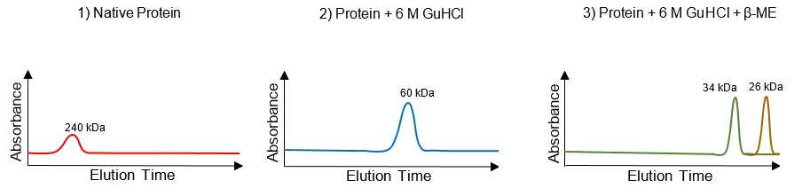
in this video, we're gonna talk about size, exclusion chromatography so size exclusion Chromatography is another type of column chromatography that's used to purify a protein based on its size, of course, and so size exclusion chromatography is also known as gel filtration chromatography, and the reason for that is because the material the stationary phase inside of the column, is made up of a gel, and it's used to filter proteins based on their size. And so we haven't yet talked about the technique gel electrophoresis. We will a little later in our course, but most of you guys have covered gel, electrophoresis and your previous courses, and so you might have a preconceived idea of how molecules are supposed to move through a gel. But I'm here to tell you guys that the way that molecules move through a gel and gel electrophoresis is a lot different than the way that molecules move through a gel in gel, filtration chromatography. And so contrary to gel electrophoresis. It's actually the larger proteins that dilute faster and earlier from the column than the smaller proteins. So the smaller proteins take longer to move through the column, and they are eluded last, And so the reason for this is actually pretty simple. And so, uh, the reason is because the stationary phase inside of the gel filtration chromatography column consists of very porous beads. And so these poorest beads have cavities in them that are engineered to be a very specific size. And different beads have different sized cavities. So let's take a look at our example down below to clear this up. And what you'll see is that gel filtration chromatography is another type of column chromatography, So notice we have our column here and inside of our column. It's packed with stationary phase, which is filled with thes poorest beads that are that have cavities engineered to be a very specific size. So if we were to zoom in on these bees, what we would see is that the beads actually have pores in them, so they have these little passageways inside of them, and some of the pores are bigger than the other ones. So some pours allow medium sized proteins go in, and other pours only allow small proteins to go in, and so notice in our protein mixture up at the top here, what we have are large proteins in red, intermediate size proteins and green and small proteins in light blue. And so notice that the light blue proteins are able toe enter all of the poorest beads, whereas the intermediate proteins can Onley enter some of them. And so notice that these pores beads, they end up taking the pores create a longer path. And so, um, if you, uh, the red proteins here, which are very large, they're actually not able to fit their physically cannot fit into the pores of these beads. So they have to take another route around the beads. They have to go around them. And so that's exactly the reason for why we see this illusion of the proteins, so large proteins actually cannot. They cannot enter the cavities of the beads because they're literally just too large to fit. And so what they're gonna do is they're going to take a shorter and faster route through the column, so these large proteins are gonna move through the column faster, and the small proteins that actually do enter the cavities of small proteins enter the cavities of the beats, and they are slowed down. They're slowed down because they have to take a longer route through the column. And so when we take a look at our column chromatography here, notice that we have our red proteins that are alluding fairly quickly and noticed that they end up coming out of the column fastest. The intermediate sized proteins, they end up coming out the second fastest, and then the slowest proteins that come out are going to be the smallest ones. And so what you'll see is that it's the largest proteins, so larger proteins are gonna be the ones that dilute from the column first, so they come out of the column first. And so if we were to measure the absorb INTs of the molecules as they come out of the bottom of the column, notice that we have the light absorbent over here, and this is a chromatic graham. So the chromatic Graham shows a light absorbent on the Y axis and the illusion time on the X axis. So how long it takes for the molecules to allude and come out of the column and notice that it's the red protein here? So the red protein has a short illusion time, so it's lower on the X axis here further to the left. And so that means that they come out of the column first and then notice that it's the green proteins that intermediate size proteins that come out of the column next. And then. Of course, it's the smallest proteins that come out of the column last, so they take the longest to come out. And so it's important to understand that column, uh, size exclusion chromatography can actually separate proteins based on their size. And also, uh, if we have proteins that we know exactly what they're sizes. We can use size exclusion chromatography to determine the size of a unknown protein, and so we'll be able to get a lot of practice with size exclusion chromatography in our next couple of videos, so I'll see you guys in those videos.
2
Problem
Problem
Which of the following statements is false?
A
In ion exchange chromatography, the bound proteins are eluted using a salt solution.
B
Gel filtration chromatography can be used to determine an unknown protein's relative molecular size/mass.
C
In gel filtration chromatography, the smallest proteins are eluted from the column last.
D
Separation of proteins in gel filtration chromatography is based on size & net charge of the proteins.
E
None of them. All above statements are true.
3
Problem
Problem
A new protein of unknown structure has been purified & gel filtration chromatography reveals that the native protein has a molecular weight of 240 kDa. Chromatography in the presence of 6 M guanidine hydrochloride (GuHCl), a chaotropic agent that has a similar effect on proteins as urea, yields a single absorbance peak corresponding to a protein of Mr 60 kDa. Chromatography in the presence both of 6 M guanidine hydrochloride and 10 mM β-mercaptoethanol (β-ME) yields peaks for proteins of Mr 34 kDa and 26 kDa. Using this data, which option best describes the structure of this protein? Hint: sketch a visual of the protein after each chemical treatment.
A
A homotetramer (4 identical 60 kDa subunits); each subunit is a heterodimer of 2 disulfide-linked chains (34 & 26 kDa).
B
A heterooctomer (8 different subunits); four subunits each of 34-kDa & 26 kDa, all held together via disulfide bonds.
C
A homodimer (2 identical 120 kDa subunits); each subunit is a a homodimer of 2 disulfide-linked chains (60 kDa each).
D
A heterotetramer (4 different subunits); each subunit is a homodimer of 2 disulfide-linked chains (60 kDa each).
4
Problem
Problem
To answer the questions A, B & C below, use the provided chart with the properties of the four proteins.
A) What is the order of elution of the proteins from a size-exclusion chromatography column?
a. A → B → C → D.
b. D → B → A → C.
c. B → D → A → C.
d. C → A → D → B.
B) Which pH is best for separating the proteins using anion-exchange chromatography?
a. pH = 6. b. pH = 7. c. pH = 8.
C) In what order would the proteins elute from the anion-exchange chromatography column?