Pearson+ LogoPearson+ Logo
Start typing, then use the up and down arrows to select an option from the list.

Biochemistry

Learn the toughest concepts covered in Biochemistry with step-by-step video tutorials and practice problems by world-class tutors

4. Protein Structure

Tertiary Structure of Protein

1
concept

Tertiary Structure of Protein

clock
2m
Play a video:
Was this helpful?
So now that we've covered the most common types of secondary structures such as Alfa Ulysses, beta sheets and beta turns and loops, we can finally talk about tertiary protein structure. So the tertiary protein structure is referring to the folded overall three dimensional shape of a protein. And so, unlike secondary structure, it turns out that tertiary structure is actually stabilized by our group interactions. So finally the are groups are involved. So remember that for all of our secondary interactions, we said that the are groups are not involved and it's all about the backbone interactions. But for tertiary interactions, it's all about the our group interactions and not the backbone interactions. So it's kind of flipped there. And so it turns out that the amino acids are groups that are far apart in sequence can actually still interact with one another in the tertiary structure because of folding of the poly peptide chain. So it's there's no requirement for the amino acid are groups to be neighbors. They could be really far apart. You could have the very first amino acid, our group and the very last amino acid our group interact with each other because of folding of the chain. And so if we take a look at, uh down below at this image, what we'll see is that we have a typical protein structure being shown. And so what you'll see is that in red, what we have are these beta strands and beta sheets and green. What we have are the Alfa Helix and the Alfa Ulysses, and then we also have these long loops. So we've got these long loops that change backbone directions, and we know that these air going to be loops. And then we also have thes other backbone changes that are abruptly changing the directions. And so these are our beta turns. And so you can see how all of these secondary structures come together to form the overall three dimensional shape of the protein and form that together they come together to help form parts of the tertiary structure. And so before we actually get to the specific interactions specific, our group interactions that characterize tertiary structure will first get some practice. And so I'll see you guys in that practice video
2
Problem

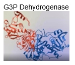
Which of the following is true regarding the tertiary structure of G3P dehydrogenase?

3
concept

Tertiary Structure Of Protein

clock
7m
Play a video:
Was this helpful?
So in our last lesson video, we said that tertiary structure is primarily stabilized by our group interactions and not by backbone interactions. And so most of these are group interactions that stabilized tertiary structure are non co Vaillant interactions. And there are actually four non co violent interactions that you guys should be familiar with. Numbered 123 and four down below. And so the first non Covalin interaction that you guys should know is ionic bonding, and Ionic bonds are also known as salt bridges. Now, the second non koval interaction is the hydrophobic effect, which we also covered in our previous lesson videos. The third is hydrogen bonding, and then the fourth is actually Vander Wal's interactions. And so, although most of the our group interaction stabilizing tertiary structure are non covalin, there are some Covalin, our group interactions that can stabilize tertiary structure. And really, that's what these next two bullet points are dedicated to. And so to Sistine amino acids can react and link to form a Sistine residue containing a single die sulfide bond. And so essentially die sulfide bonds are our fifth, uh, type of our group interaction. But this is a co Vaillant type of our group interaction. And so, uh, die sulfide bridges are a type of CO Vaillant, our group interaction that could potentially stabilize the overall three D structure or the tertiary structure of a protein. And so again, all of these bonds here are numbered. 1234 and five. And the numbers up above in the tax correspond with numbers that you see down below and our image. And so in our image below, we're gonna talk about on give examples of each of these types of bonds. And so first, which will notice, is this long, orange looking rope thing right here is essentially our peptide backbone, and so notice that the peptide backbone is capable of folding, so notice that it's not linear. It's actually fold into different patterns. And so we're going to talk more about protein folding a little later in our course. But it's important to note that the protein folding allows for amino acids that are distant in sequence to be put closer into proximity so that there are groups can interact, and we'll be able to see that down below as well. And so we know that our peptide backbone has an end terminal end and a C terminal and on the opposite side. And so our first non covalin interaction is actually the Ionic Bond or Ionic bonding. And we know that Ionic bonds are also known as salt bridges, and so Ionic bonds can Onley form between our groups that are ionized able and recall that are demonic for memorizing the seven amino acids with ionized able are groups is just yucky crazy dragons eat nights riding horses. And so these are the seven amino acids that are ionized able, and those are the ones that are capable of forming ionic bonds. And so, in this example here, you can see that we have, uh, to ionized able are groups We have license our group right here and then on the other side, what we have is ah, Spartak acids are group, and so a Spartak acid is d here for the one letter coat and listen is K. And so our second type of non covalin interaction is the hydrophobic effect, and so we can see the hydrophobic effect occurring between these two non polar amino acids. More specifically, these two veiling amino acids and so the hydrophobic effect essentially allows these two non polar veiling amino acid are groups to clump together, as we see here now, our third non covalin interaction is the hydrogen bond. And so hydrogen bonding can really occur between any amino acid, our group that conform and ah, hydrogen bond but typically will see that these are the polar amino acids are groups. And so here you can see that we have a Syrian, our group right here, which is one of our polar amino acids. And then we also have a sparrow. Genes are group, and both of these our groups are polar, and they're forming a hydrogen bond with one another. And so notice that we do not actually have 1/4 number for the Vander wal's interactions. And that's because the Vander Waals interactions occurs between all atoms. And so the Vander Waals interactions are occurring between all portions of our Polly peptide chain. And you can imagine that the Vander Waals interactions are occurring between these two separate chains here to keep them in close proximity, and overall, they have a large impact on, uh providing tertiary structure to our protein. But because they occur throughout our entire chain. We don't have them numbered. And they one particular point. Now, our fifth and final type of our group interaction is actually a co Vaillant, our group interaction, and that is our die sulfide bridge. And so you can see that the dye sulfide bridge essentially forms between two Sistine amino acids. Toe form the dye sulfide bond that's shown in red between these two solvers. And we can see that a little bit better over here on the image on the right. And so you can see what we have are two Sistine amino acids on the left hand portion over here so you can see here's one Sistine Amino acid and here's the other. And recall that Sistine spends ah lot of time in the Sistine Chapel. And whenever somebody makes a noise, systems like So, Sistine is really just a winning within uh S H Group, A group coming off. And so that's how we can remember Sistine, our group. And so here we have 1 16. Here we have another, and when they are in close enough proximity, they're able to react to form a single Sistine Residue, just like what we said. Up above a single Sistine residue can be form, and this one single Sistine residue contains a dice sulfide bridge and the dice sulfide bridge is in red between these two solvers right here. And so essentially, you can see that although most of our our group interactions are non covalin, we do have die sulfide bridges that are a type of covalin, our group interaction. And together all five of these are group interactions contribute to the tertiary structure off proteins. And so this concludes our lesson on the tertiary structure of proteins and the our group interactions, and we'll be able to get some practice in our next couple of videos, so I'll see you guys there.
4
Problem

True or False: At pH 2, Gln & Met R-groups can form a salt bridge to stabilize tertiary structure.

5
Problem

Which statement regarding the tertiary structure of Ribonuclease A interactions is true?

Divider