Assume you stain Bacillus by applying malachite green with heat and then counterstain with safranin. Through the microscope, the green structures are a. Cell walls b. Capsules c. Endospores d. Flagella e. Impossible to identify
Verified step by step guidance
1
Understand the staining procedure described: malachite green is applied with heat, followed by a counterstain with safranin. This is a classic method used in microbiology known as the endospore stain.
Recall that malachite green is a primary stain that penetrates tough structures like endospores when heat is applied, allowing the dye to enter and bind inside the endospore.
Recognize that after staining with malachite green, the cells are counterstained with safranin, which stains the vegetative cells red or pink, while the endospores retain the green color because they resist decolorization.
Identify that the green structures observed under the microscope correspond to the endospores, as they are the only structures that retain the malachite green stain after the counterstain.
Conclude that the correct identification of the green structures stained by malachite green with heat and counterstained with safranin is endospores.
Verified video answer for a similar problem:
This video solution was recommended by our tutors as helpful for the problem above
Video duration:
1m
Play a video:
0 Comments
Key Concepts
Here are the essential concepts you must grasp in order to answer the question correctly.
Endospore Staining Technique
The endospore stain uses malachite green as the primary stain applied with heat to penetrate the tough outer layer of endospores. Heat acts as a mordant, allowing the dye to enter and bind to the endospore. After staining, a counterstain like safranin colors the vegetative cells, making endospores appear green under the microscope.
Endospores are highly resistant, dormant structures formed by some bacteria like Bacillus to survive harsh conditions. They have a tough outer coat that resists staining and environmental damage. Their unique composition requires special staining methods to visualize them distinctly from vegetative cells.
Counterstaining involves applying a second stain, such as safranin, after the primary stain to color cells or structures that do not retain the first dye. In endospore staining, this step colors the vegetative cells red or pink, providing contrast to the green-stained endospores and allowing clear differentiation under the microscope.

 Verified step by step guidance
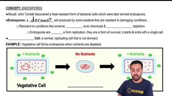
 05:35
05:35