Indicate which form of electron microscopy is being described: SEM, TEM, or both. a. Images the external surface of a specimen b. Sample must be thinly sliced prior to viewing c. Provides details about internal structures of the sample d. Can be used to image viruses e. Provides black-and-white images f. Specimen is dead
Verified step by step guidance
1
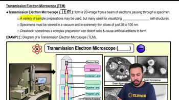
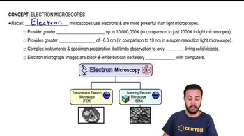
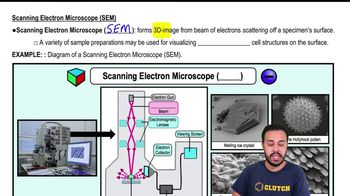
Understand the two main types of electron microscopy: Scanning Electron Microscopy (SEM) and Transmission Electron Microscopy (TEM). SEM images the surface of specimens by scanning with electrons, while TEM transmits electrons through thinly sliced specimens to reveal internal structures.
For each statement, determine which microscopy technique it describes based on the characteristics: (a) SEM images external surfaces, so it corresponds to SEM.
(b) Since TEM requires electrons to pass through the sample, the specimen must be thinly sliced, so this describes TEM.
(c) Providing details about internal structures is a hallmark of TEM, as it allows visualization inside the specimen.
(d) Both SEM and TEM can be used to image viruses, but TEM is more commonly used due to its higher resolution for internal viral structures, so this can be both or primarily TEM.
(e) Both SEM and TEM produce black-and-white images because electron microscopes detect electrons rather than light, so this applies to both.
(f) Specimens must be dead for both SEM and TEM because the preparation process involves vacuum and electron beam exposure, which are incompatible with living samples.
Verified video answer for a similar problem:
This video solution was recommended by our tutors as helpful for the problem above.
Video duration:
4m
Play a video:
0 Comments
Key Concepts
Here are the essential concepts you must grasp in order to answer the question correctly.
Scanning Electron Microscopy (SEM)
SEM produces detailed images of the external surface of specimens by scanning with a focused electron beam. It provides three-dimensional-like images and requires specimens to be coated with a conductive material. SEM is ideal for studying surface morphology but does not reveal internal structures.
TEM transmits electrons through ultra-thin slices of a specimen to visualize internal structures at very high resolution. Samples must be thinly sliced and often stained with heavy metals to enhance contrast. TEM is commonly used to study cellular organelles and viruses.
Specimen Preparation and Imaging Characteristics in Electron Microscopy
Both SEM and TEM require specimens to be dead and prepared carefully, often involving fixation and dehydration. Electron microscopy images are typically black-and-white due to electron detection methods. Both techniques can image viruses, but TEM is more commonly used for internal viral structure analysis.

 4:52 minutes
 Verified step by step guidance
 02:42
02:42